. 2018 Aug 28;8(1):12983.
doi: 10.1038/s41598-018-31214-x.

Dynamic expression of HOPX in alveolar epithelial cells reflects injury and repair during the progression of pulmonary fibrosis

Affiliations

Dynamic expression of HOPX in alveolar epithelial cells reflects injury and repair during the progression of pulmonary fibrosis

Chiharu Ota et al. Sci Rep. .

Abstract

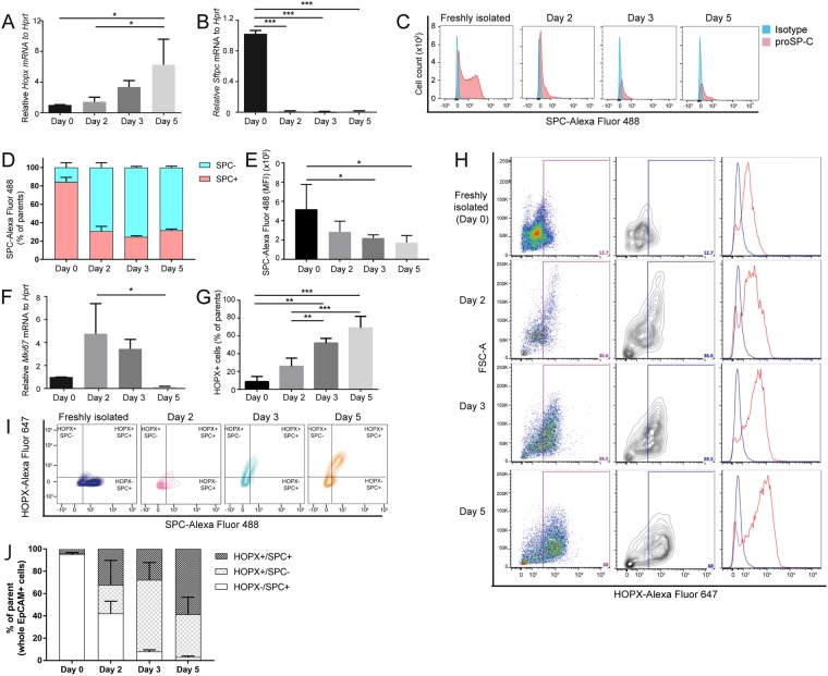
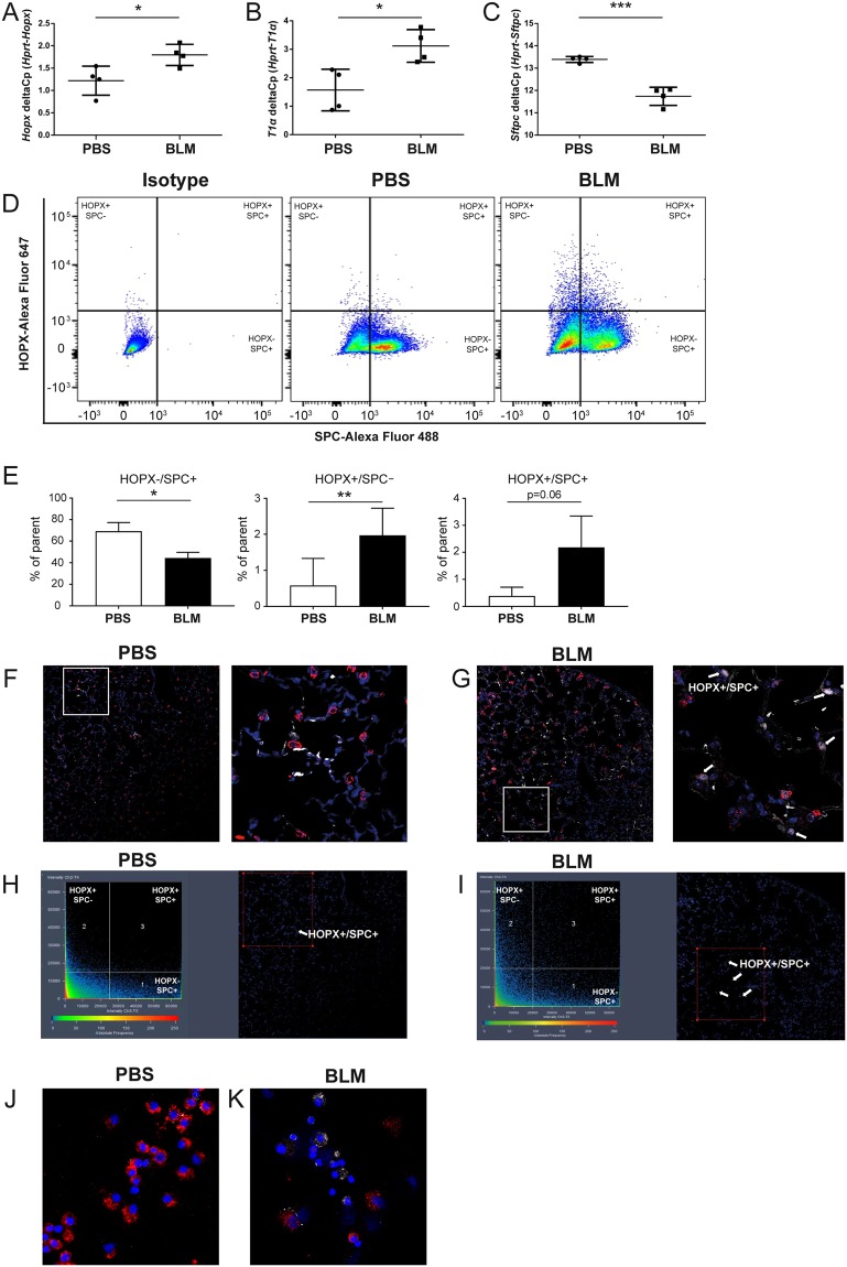
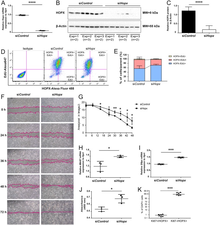
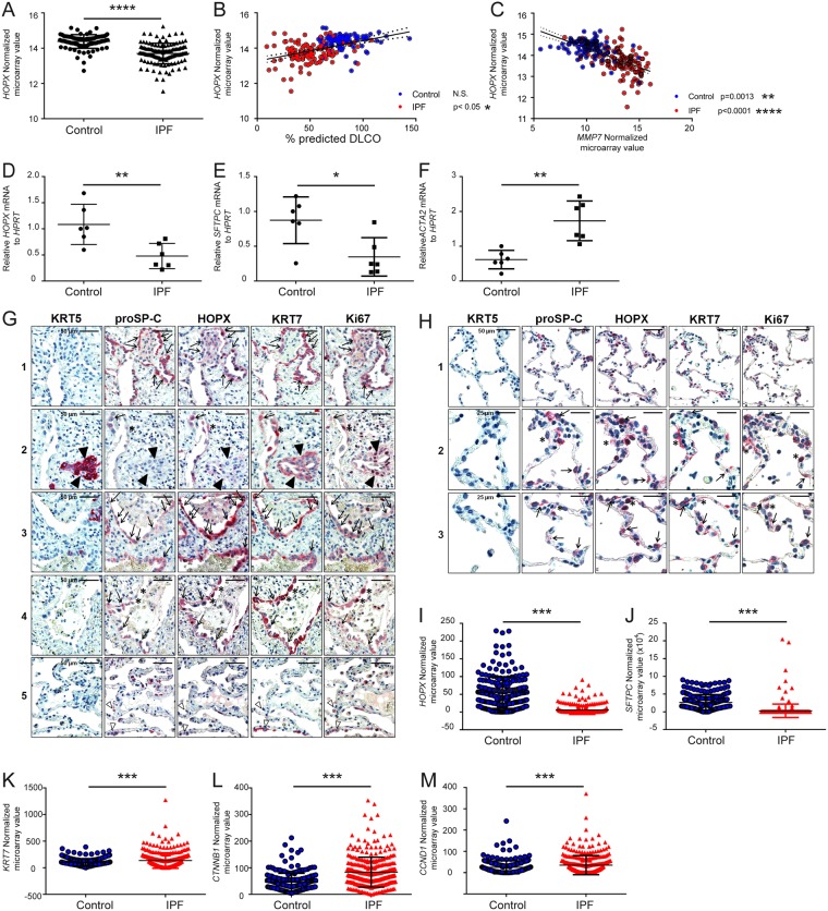
Mechanisms of injury and repair in alveolar epithelial cells (AECs) are critically involved in the progression of various lung diseases including idiopathic pulmonary fibrosis (IPF). Homeobox only protein x (HOPX) contributes to the formation of distal lung during development. In adult lung, alveolar epithelial type (AT) I cells express HOPX and lineage-labeled Hopx+ cells give rise to both ATI and ATII cells after pneumonectomy. However, the cell function of HOPX-expressing cells in adult fibrotic lung diseases has not been investigated. In this study, we have established a flow cytometry-based method to evaluate HOPX-expressing cells in the lung. HOPX expression in cultured ATII cells increased over culture time, which was accompanied by a decrease of proSP-C, an ATII marker. Moreover, HOPX expression was increased in AECs from bleomycin-instilled mouse lungs in vivo. Small interfering RNA-based knockdown of Hopx resulted in suppressing ATII-ATI trans-differentiation and activating cellular proliferation in vitro. In IPF lungs, HOPX expression was decreased in whole lungs and significantly correlated to a decline in lung function and progression of IPF. In conclusion, HOPX is upregulated during early alveolar injury and repair process in the lung. Decreased HOPX expression might contribute to failed regenerative processes in end-stage IPF lungs.


Conflict of interest statement

The authors declare no competing interests.

Figures

Figure 1
Figure 1
Expression of HOPX and proSP-C during ATII-ATI cell trans-differentiation in vitro. Quantitative (q) RT-PCR analysis of (A) Hopx (B) Sftpc, (C,D) FCM analysis of proSP-C expression over culture, (E) mean fluorescent intensity (MFI) of proSP-C-Alexa-fluor 488, and (F) qRT-PCR analysis of Mki67 during 5 days culture of pmATII cells (n = 3). (G,H) FCM-based quantification of HOPX expression during the culture. (I) and (J) FCM-based quantification of HOPX/proSP-C co-expression during the culture (n = 3). *p < 0.05, **p < 0.01, ***p < 0.005.
Figure 2
Figure 2
Expression of HOPX/proSP-C lung epithelial cell subpopulations in BLM-induced pulmonary fibrosis model in vivo. Quantitative RT-PCR analysis of (A) Hopx (B) T1α, and (C) Sftpc in EpCAM+ cells from PBS or BLM-instilled lungs (n = 4). (D) FCM-based evaluation of HOPX/proSP-C expression in pmATII cells from PBS or BLM-instilled lungs (representative images from n = 3). Quantification of (E) HOPX−/proSP-C+, HOPX+/proSP-C−, and HOPX+/proSP-C+ cells in PBS and BLM-instilled lungs (n = 3). Immunofluorescence staining (IF) of HOPX (white) and proSP-C (red) in the sections from (F) PBS and (G) BLM-instilled lungs. Visualization of the co-expression of HOPX and proSP-C from (H) PBS and (I) BLM-instilled lungs using ZEN2009 software. IF of HOPX (white) and proSP-C (red) in cytospun EpCAM+ cells from (J) PBS and (K) BLM-instilled lungs. *p < 0.05, **p < 0.01, ***p < 0.005.
Figure 3
Figure 3
Effect of HOPX on proliferation and differentiation in MLE12 epithelial cells in vitro. Evaluation of si-RNA-based Hopx knockdown in MLE12 lung epithelial cells with (A) qRT-PCR of Hopx, (B) Western blotting of HOPX, (C) quantification of Western blotting data. (D) EdU assay of MLE12 lung epithelial cells co-stained with HOPX antibody in vitro. Alexa Fluor 647-EdU+ cells and Alexa Fluor 488-HOPX+ cells were evaluated by FCM (Representative image of n = 2). (E) Quantification of HOPX+ EdU+ cells and HOPX-EdU+ cells in MLE12 cells (n = 2). (F,G) Cellular scratch assay, qRT-PCR analysis of (H) Mki67 and (I) Sftpc, and (J) WST assay of MLE12 cells transfected with control siRNA and Hopx siRNA. (J) The ratio of Ki67 positive/negative cells with HOPX co-expression in IF. *p < 0.05, **p < 0.01, ***p < 0.005.
Figure 4
Figure 4
Expression and localization of HOPX in control (donor) and IPF lungs. In silico analysis from Lung Genomics Research Consortium (LGRC) of (A) HOPX mRNA expression in organ donor (control) and IPF lungs, (B) correlation between HOPX mRNA expression and DLco (blue dots: control, red dots: IPF). (C) Correlation between HOPX mRNA and MMP7 mRNA in control (blue dots) and IPF (red dots) in silico. QRT-PCR analysis of (D) HOPX mRNA, (E) SFTPC mRNA, and (F) ACTA2 mRNA from whole lung homogenate of control and IPF lungs in our cohort. (G) Immunohistochemical staining of serial sections of control and IPF lung tissue for KRT5, proSP-C, HOPX, KRT7, and Ki67. In IPF lungs, proSP-C+/KRT7+ cells revealed cytoplasmic or in part nuclear expression of HOPX without nuclear Ki67 (Indicated by arrows in panels 1–4). ProSP-Clow/KRT7+ AECs of IPF-lungs also indicated robust HOPX-immunoreactivity with nuclear Ki67 expression (asterisks in panel 2 and 4). Some proSP-C+/KRT7+ cells without HOPX immunoreactivity were observed in the area of relatively preserved alveolar septa (white arrowheads in panel 5). KRT5+/KRT7+ bronchiolar basal cells did not express HOPX, and indicated nuclear expression of Ki67 (Indicated by black arrowheads in panel 2). (H) Immunohistochemical staining of serial sections of control donor lung tissues for KRT5, proSP-C, HOPX, KRT7, and Ki67. In donor lungs, HOPX expression was present in proSP-C+/KRT7+ ATII (arrows) and proSP-Clow/KRT7+ AECs (asterisks) (panels 2 and 3 in Fig. 4O), and faint in normal bronchiolar epithelium. ProSP-Clow/KRT7+ AECs expressing HOPX also revealed nuclear Ki67 expression (indicated by asterisks). In silico single cell RNA sequence analysis of (I) HOPX, (J) SFTPC, (K) KRT7, (L) CTNNB1, and (M) CTND1 in EpCAM+/HTII280+ cells. *p < 0.05, **p < 0.01, ***p < 0.005, ****p < 0.0001.

Similar articles

Cited by

References

    1. Knudsen L, Ruppert C, Ochs M. Tissue remodelling in pulmonary fibrosis. Cell Tissue Res. 2017;367:607–626. doi: 10.1007/s00441-016-2543-2. - DOI - PubMed
    1. Williams MC. Alveolar type I cells: molecular phenotype and development. Annu Rev Physiol. 2003;65:669–695. doi: 10.1146/annurev.physiol.65.092101.142446. - DOI - PubMed
    1. Ward HE, Nicholas TE. Alveolar type I and type II cells. Aust N Z J Med. 1984;14:731–734. doi: 10.1111/j.1445-5994.1984.tb04343.x. - DOI - PubMed
    1. Kuroki Y, Voelker DR. Pulmonary surfactant proteins. J Biol Chem. 1994;269:25943–25946. - PubMed
    1. Fehrenbach H. Alveolar epithelial type II cell: defender of the alveolus revisited. Respir Res. 2001;2:33–46. doi: 10.1186/rr36. - DOI - PMC - PubMed

Publication types

MeSH terms