

Generation of customized orbital implant templates using 3-dimensional printing for orbital wall reconstruction
- PMID: 30154573
- PMCID: PMC6293000
- DOI: 10.1038/s41433-018-0193-1
Generation of customized orbital implant templates using 3-dimensional printing for orbital wall reconstruction
Abstract
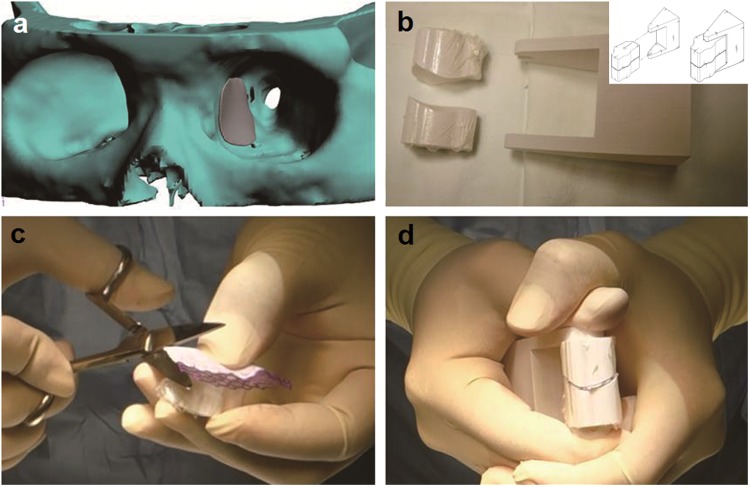
Objectives: To describe and evaluate a novel surgical approach to orbital wall reconstruction that uses three-dimensionally (3D) printed templates to mold a customized orbital implant.
Methods: A review was conducted of 11 consecutive patients who underwent orbital wall reconstruction using 3D-printed customized orbital implant templates. In these procedures, the orbital implant was 3D pressed during surgery and inserted into the fracture site. The outcomes of this approach were analyzed quantitatively by measuring the orbital tissue volumes within the bony orbit using computed tomography.
Results: All 11 orbital wall reconstructions (6 orbital floor and 5 medial wall fractures) were successful with no post operative ophthalmic complications. Statistically significant differences were found between the preoperative and post operative orbital tissue volumes for the affected orbit (24.00 ± 1.74 vs 22.31 ± 1.90 cm3; P = 0.003). There was no statistically significant difference found between the tissue volume of the contralateral unaffected orbit and the affected orbit after reconstruction (22.01 ± 1.60 cm3 vs 22.31 ± 1.90 cm3; P = 0.182).
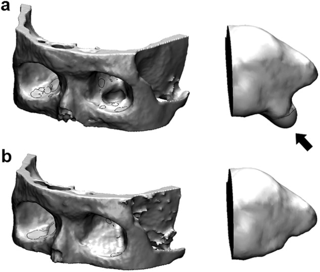
Conclusion: 3D-printed customized orbital implant templates can be used to press and trim conventional implantable materials with patient-specific contours and sizes for optimal orbital wall reconstruction. It is difficult to design an orbital implant that exactly matches the shape and surface of a blowout fracture site due to the unique 3D structure of the orbit. The traditional surgical method is to visually inspect the fracture site and use eye measurements to cut a two-dimensional orbital implant that corresponds to the anatomical structure of the fracture site. However, implants that do not fit the anatomical structure of a fracture site well can cause complications such as enophthalmos, diplopia and displacement of the implant.
Conflict of interest statement
The authors declare that they have no conflict of interest.
Figures

Similar articles
-
Customized Titanium Mesh Based on the 3D Printed Model vs. Manual Intraoperative Bending of Titanium Mesh for Reconstructing of Orbital Bone Fracture: A Randomized Clinical Trial.Rev Recent Clin Trials. 2017;12(3):154-158. doi: 10.2174/1574887112666170821165206. Rev Recent Clin Trials. 2017. PMID: 28828975 Clinical Trial.
-
Technical concept of patient-specific, ultrahigh molecular weight polyethylene orbital wall implant.J Craniomaxillofac Surg. 2013 Jun;41(4):282-90. doi: 10.1016/j.jcms.2012.10.007. Epub 2013 Jan 18. J Craniomaxillofac Surg. 2013. PMID: 23333489
-
Customized Orbital Wall Reconstruction Using Three-Dimensionally Printed Rapid Prototype Model in Patients With Orbital Wall Fracture.J Craniofac Surg. 2016 Nov;27(8):2020-2024. doi: 10.1097/SCS.0000000000003195. J Craniofac Surg. 2016. PMID: 28005746
-
Three-dimensional (3D) printing for post-traumatic orbital reconstruction, a systematic review and meta-analysis.Br J Oral Maxillofac Surg. 2022 Nov;60(9):1176-1183. doi: 10.1016/j.bjoms.2022.07.001. Epub 2022 Jul 16. Br J Oral Maxillofac Surg. 2022. PMID: 35931592
-
Patient-specific Implants for Orbital Fractures: A Systematic Review.Ophthalmic Plast Reconstr Surg. 2022 Sep-Oct 01;38(5):417-424. doi: 10.1097/IOP.0000000000002089. Epub 2021 Nov 8. Ophthalmic Plast Reconstr Surg. 2022. PMID: 34750315
Cited by
-
Theoretical model of pediatric orbital trapdoor fractures and provisional personalized 3D printing-assisted surgical solution.Bioact Mater. 2020 Sep 14;6(2):559-567. doi: 10.1016/j.bioactmat.2020.08.029. eCollection 2021 Feb. Bioact Mater. 2020. PMID: 33005822 Free PMC article.
-
Investigating the accuracy of mandibulectomy and reconstructive surgery using 3D customized implants and surgical guides in a rabbit model.Maxillofac Plast Reconstr Surg. 2023 Jan 26;45(1):8. doi: 10.1186/s40902-023-00375-9. Maxillofac Plast Reconstr Surg. 2023. PMID: 36701071 Free PMC article.
-
Preoperative 3D Reconstruction Model in Slow Mohs Surgery for Dermatofibrosarcoma Protuberans.Comput Intell Neurosci. 2022 Apr 7;2022:5509129. doi: 10.1155/2022/5509129. eCollection 2022. Comput Intell Neurosci. 2022. Retraction in: Comput Intell Neurosci. 2023 Dec 13;2023:9854361. doi: 10.1155/2023/9854361. PMID: 35432518 Free PMC article. Retracted. Review.
-
3D Printing in Eye Care.Ophthalmol Ther. 2021 Dec;10(4):733-752. doi: 10.1007/s40123-021-00379-6. Epub 2021 Jul 29. Ophthalmol Ther. 2021. PMID: 34327669 Free PMC article. Review.
-
Rehearsal simulation to determine the size of device for left atrial appendage occlusion using patient-specific 3D-printed phantoms.Sci Rep. 2022 May 11;12(1):7746. doi: 10.1038/s41598-022-11967-2. Sci Rep. 2022. PMID: 35546178 Free PMC article.
References
MeSH terms
LinkOut - more resources
Full Text Sources
Other Literature Sources
Medical
