

Larger Than Life: Isolation and Genomic Characterization of a Jumbo Phage That Infects the Bacterial Plant Pathogen, Agrobacterium tumefaciens
- PMID: 30154772
- PMCID: PMC6102473
- DOI: 10.3389/fmicb.2018.01861
Larger Than Life: Isolation and Genomic Characterization of a Jumbo Phage That Infects the Bacterial Plant Pathogen, Agrobacterium tumefaciens
Abstract
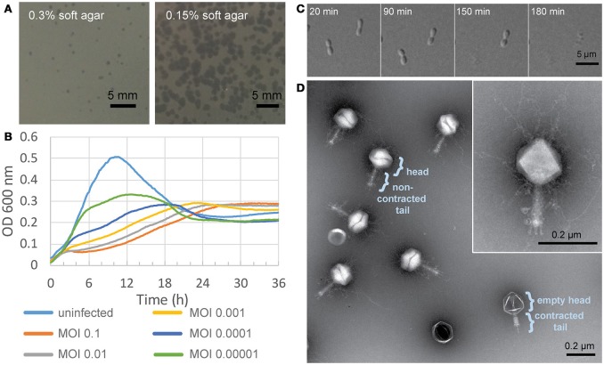
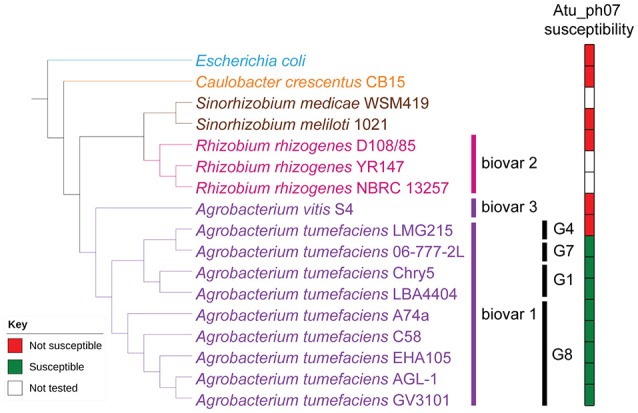
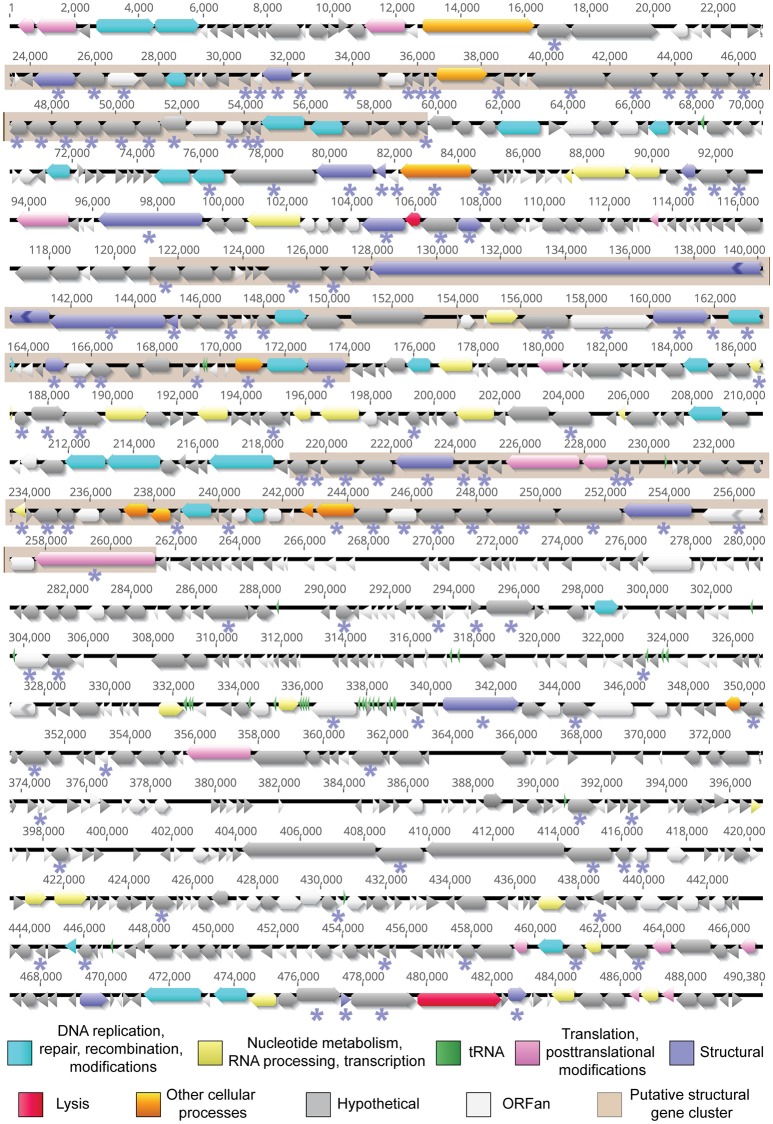
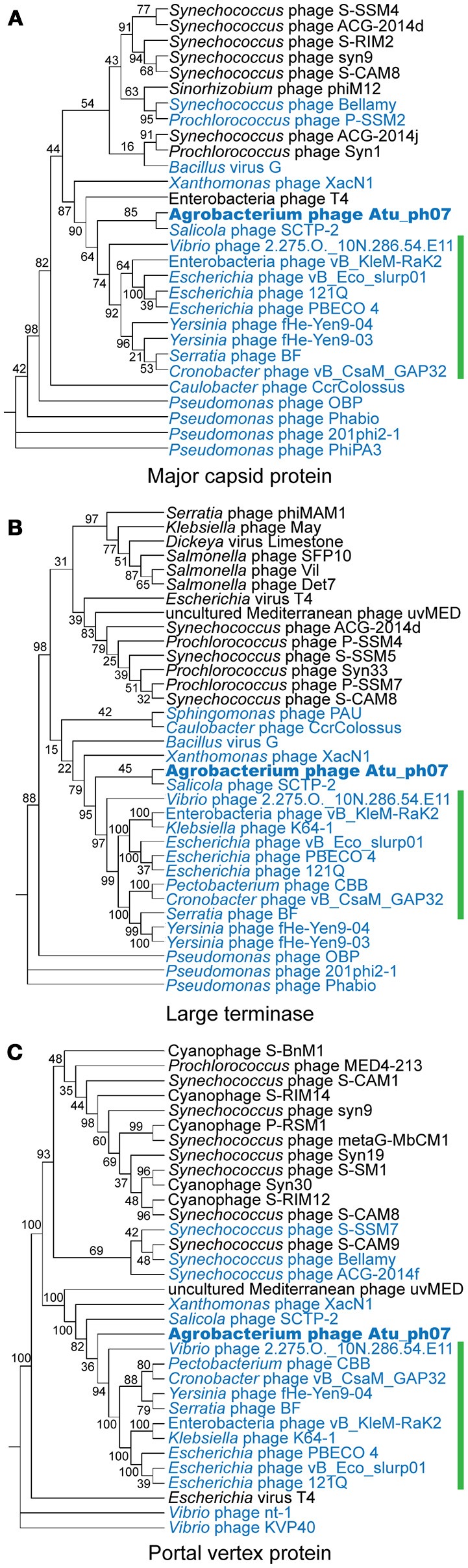
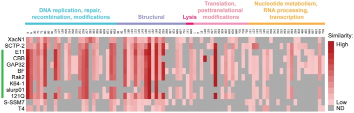
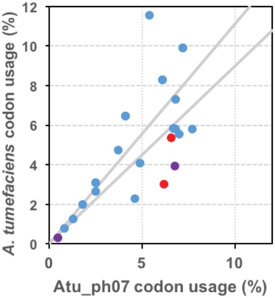
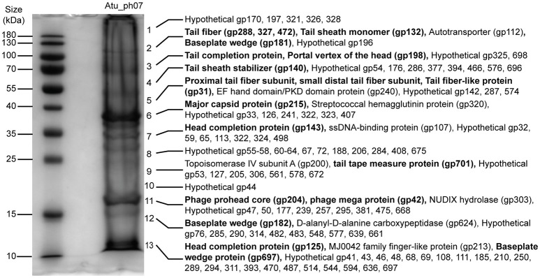
Agrobacterium tumefaciens is a plant pathogen that causes crown gall disease, leading to the damage of agriculturally-important crops. As part of an effort to discover new phages that can potentially be used as biocontrol agents to prevent crown gall disease, we isolated and characterized phage Atu_ph07 from Sawyer Creek in Springfield, MO, using the virulent Agrobacterium tumefaciens strain C58 as a host. After surveying its host range, we found that Atu_ph07 exclusively infects Agrobacterium tumefaciens. Time-lapse microscopy of A. tumefaciens cells subjected to infection at a multiplicity of infection (MOI) of 10 with Atu_ph07 reveals that lysis occurs within 3 h. Transmission electron microscopy (TEM) of virions shows that Atu_ph07 has a typical Myoviridae morphology with an icosahedral head, long tail, and tail fibers. The sequenced genome of Atu_ph07 is 490 kbp, defining it as a jumbo phage. The Atu_ph07 genome contains 714 open reading frames (ORFs), including 390 ORFs with no discernable homologs in other lineages (ORFans), 214 predicted conserved hypothetical proteins with no assigned function, and 110 predicted proteins with a functional annotation based on similarity to conserved proteins. The proteins with predicted functional annotations share sequence similarity with proteins from bacteriophages and bacteria. The functionally annotated genes are predicted to encode DNA replication proteins, structural proteins, lysis proteins, proteins involved in nucleotide metabolism, and tRNAs. Characterization of the gene products reveals that Atu_ph07 encodes homologs of 16 T4 core proteins and is closely related to Rak2-like phages. Using ESI-MS/MS, the majority of predicted structural proteins could be experimentally confirmed and 112 additional virion-associated proteins were identified. The genomic characterization of Atu_ph07 suggests that this phage is lytic and the dynamics of Atu_ph07 interaction with its host indicate that this phage may be suitable for inclusion in a phage cocktail to be used as a biocontrol agent.
Keywords: Agrobacterium; Atu_ph07; biocontrol; genomics; jumbo bacteriophage; mass spectrometry.
Figures
Similar articles
-
Isolation and Characterization T4- and T7-Like Phages that Infect the Bacterial Plant Pathogen Agrobacterium tumefaciens.Viruses. 2019 Jun 7;11(6):528. doi: 10.3390/v11060528. Viruses. 2019. PMID: 31181591 Free PMC article.
-
Expression of a Peptidoglycan Hydrolase from Lytic Bacteriophages Atu_ph02 and Atu_ph03 Triggers Lysis of Agrobacterium tumefaciens.Appl Environ Microbiol. 2017 Nov 16;83(23):e01498-17. doi: 10.1128/AEM.01498-17. Print 2017 Dec 1. Appl Environ Microbiol. 2017. PMID: 28970228 Free PMC article.
-
Soil Giant Phage: Genome and Biological Characteristics of Sinorhizobium Jumbo Phage.Int J Mol Sci. 2024 Jul 5;25(13):7388. doi: 10.3390/ijms25137388. Int J Mol Sci. 2024. PMID: 39000497 Free PMC article.
-
Intracellular Organization by Jumbo Bacteriophages.J Bacteriol. 2020 Dec 18;203(2):e00362-20. doi: 10.1128/JB.00362-20. Print 2020 Dec 18. J Bacteriol. 2020. PMID: 32868402 Free PMC article. Review.
-
The Phage Nucleus and PhuZ Spindle: Defining Features of the Subcellular Organization and Speciation of Nucleus-Forming Jumbo Phages.Front Microbiol. 2021 Jul 13;12:641317. doi: 10.3389/fmicb.2021.641317. eCollection 2021. Front Microbiol. 2021. PMID: 34326818 Free PMC article. Review.
Cited by
-
Phage Therapy for Crops: Concepts, Experimental and Bioinformatics Approaches to Direct Its Application.Int J Mol Sci. 2022 Dec 25;24(1):325. doi: 10.3390/ijms24010325. Int J Mol Sci. 2022. PMID: 36613768 Free PMC article. Review.
-
Jumbo Phages: A Comparative Genomic Overview of Core Functions and Adaptions for Biological Conflicts.Viruses. 2021 Jan 5;13(1):63. doi: 10.3390/v13010063. Viruses. 2021. PMID: 33466489 Free PMC article.
-
Characterization and genomic analysis of Sharanji: a jumbo bacteriophage of Escherichia coli.Virol J. 2025 Mar 10;22(1):67. doi: 10.1186/s12985-025-02646-5. Virol J. 2025. PMID: 40065321 Free PMC article.
-
Identification of Huge Phages from Wastewater Metagenomes.Viruses. 2023 Nov 28;15(12):2330. doi: 10.3390/v15122330. Viruses. 2023. PMID: 38140571 Free PMC article.
-
The chirality of imazethapyr herbicide selectively affects the bacterial community in soybean field soil.Environ Sci Pollut Res Int. 2019 Jan;26(3):2531-2546. doi: 10.1007/s11356-018-3736-x. Epub 2018 Nov 24. Environ Sci Pollut Res Int. 2019. PMID: 30474807
References
Grants and funding
LinkOut - more resources
Full Text Sources
Other Literature Sources
Research Materials