. 2018 Aug 1:2018:7578965.
doi: 10.1155/2018/7578965. eCollection 2018.

Luminescent Iridium Complex-Peptide Hybrids (IPHs) for Therapeutics of Cancer: Design and Synthesis of IPHs for Detection of Cancer Cells and Induction of Their Necrosis-Type Cell Death

Affiliations

Luminescent Iridium Complex-Peptide Hybrids (IPHs) for Therapeutics of Cancer: Design and Synthesis of IPHs for Detection of Cancer Cells and Induction of Their Necrosis-Type Cell Death

Abdullah-Al Masum et al. Bioinorg Chem Appl. .

Abstract

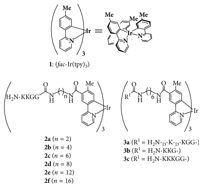
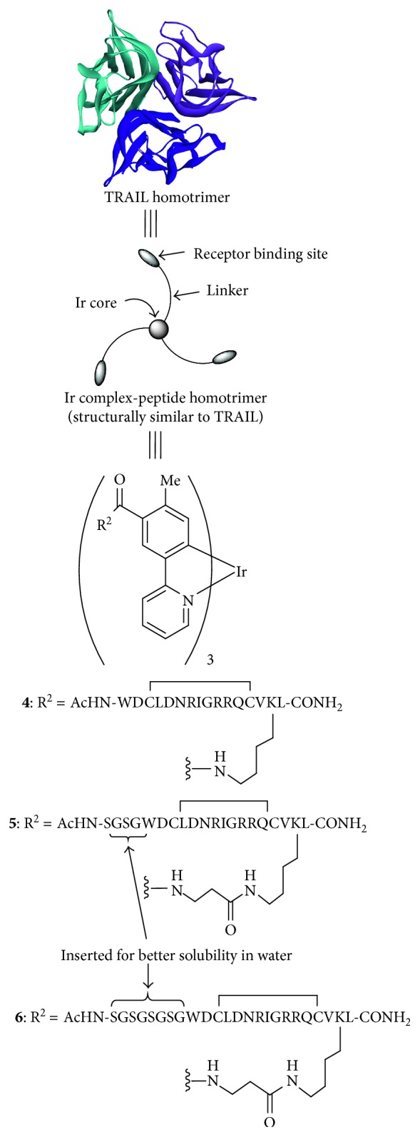
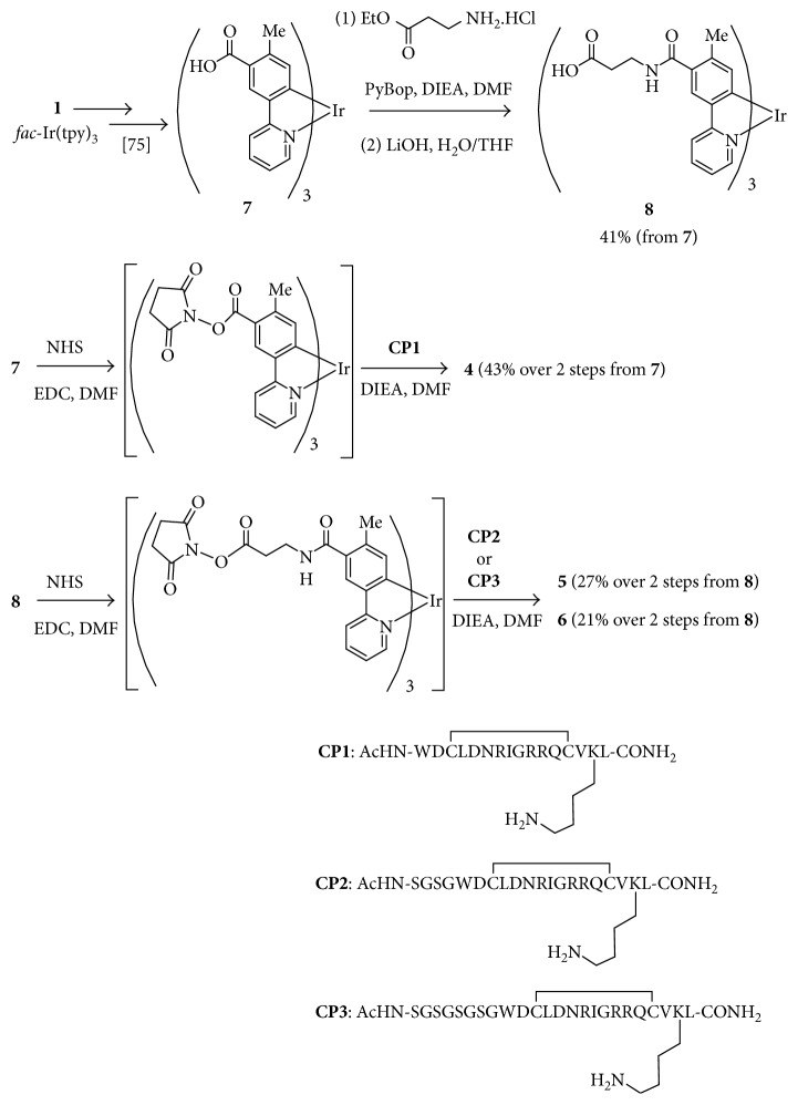
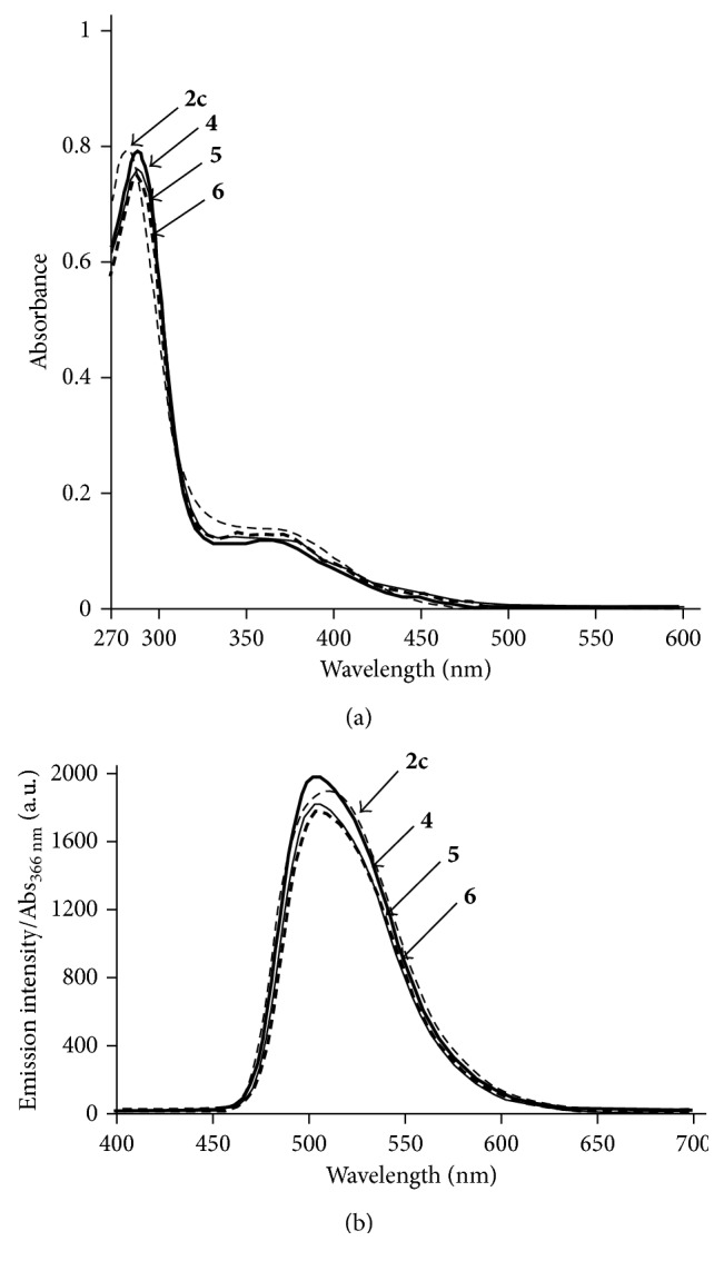
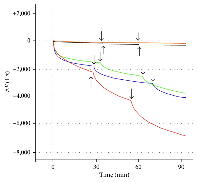
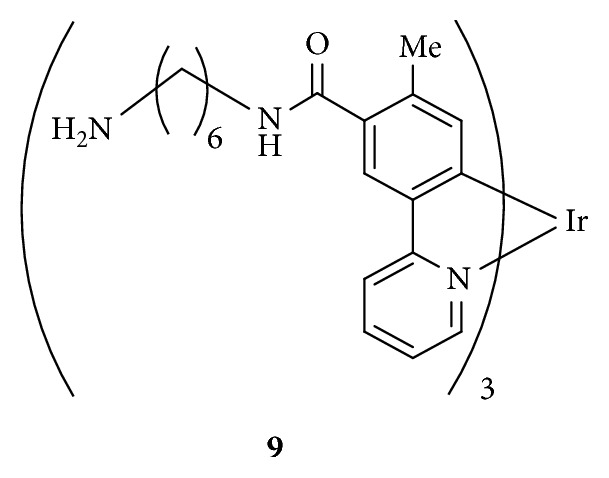
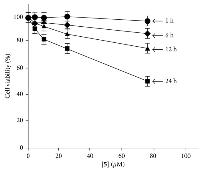
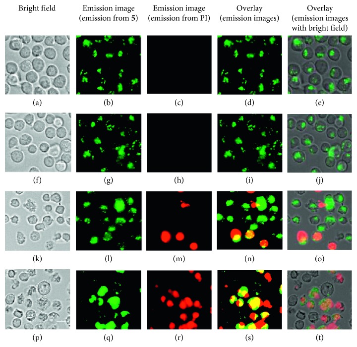
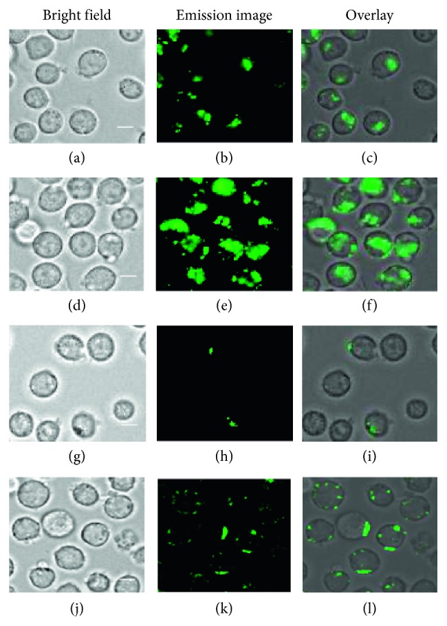
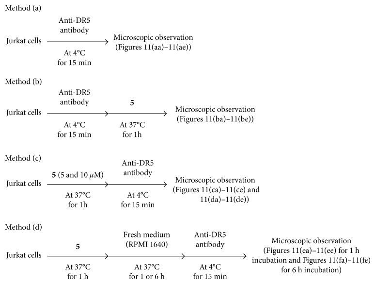
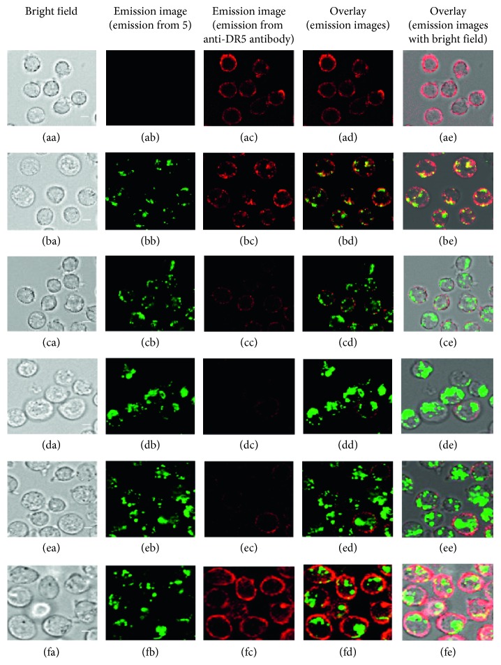
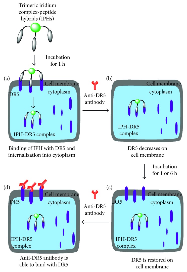
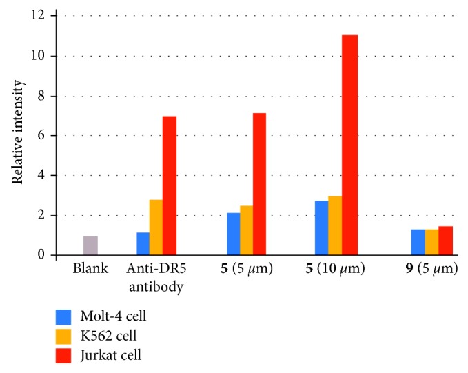
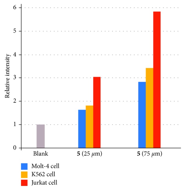
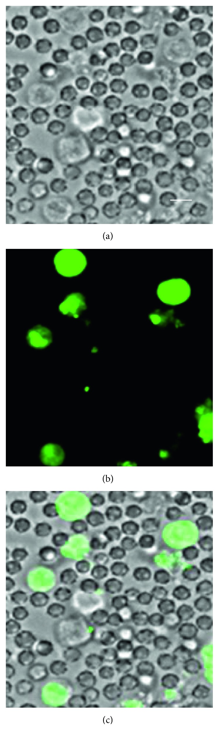
Death receptors (DR4 and DR5) offer attractive targets for cancer treatment because cancer cell death can be induced by apoptotic signal upon binding of death ligands such as tumor necrosis factor-related apoptosis-inducing ligand (TRAIL) with death receptors. Cyclometalated iridium(III) complexes such as fac-Ir(tpy)3 (tpy = 2-(4-tolyl)pyridine) possess a C3-symmetric structure like TRAIL and exhibit excellent luminescence properties. Therefore, cyclometalated Ir complexes functionalized with DR-binding peptide motifs would be potent TRAIL mimics to detect cancer cells and induce their cell death. In this study, we report on the design and synthesis of C3-symmetric and luminescent Ir complex-peptide hybrids (IPHs), which possess cyclic peptide that had been reported to bind DR5. The results of 27 MHz quartz-crystal microbalance (QCM) measurements of DR5 with IPHs and costaining experiments of IPHs and anti-DR5 antibody, suggest that IPHs bind with DR5 and undergo internalization into cytoplasm, possibly via endocytosis. It was also found that IPHs induce slow cell death of these cancer cells in a parallel manner to the DR5 expression level. These results indicate that IPHs may offer a promising tool as artificial luminescent mimics of death ligands to develop a new category of anticancer agents that detect and kill cancer cells.


Figures

Figure 1
Figure 1
Ir complexes having cationic peptides.
Figure 2
Figure 2
C 3-symmetric tris-cyclometalated Ir complex-peptide hybrids (IPHs).
Figure 3
Figure 3
Synthesis of the Ir complex-peptide hybrids (IPHs).
Figure 4
Figure 4
UV/Vis spectra of (a) 2c (dashed curve), 4 (bold curve), 5 (plain curve), and 6 (bold dashed curve). Emission spectra of (b) 2c (dashed curve), 4 (bold curve), 5 (plain curve), and 6 (bold dashed curve), in degassed DMSO at 25°C ([Ir complex] = 10 μM, excitation at 366 nm) (a.u. is the arbitrary unit).
Figure 5
Figure 5
Time course of frequency change (ΔF (Hz)) of IPHs-DR5 complexation. Conditions: temperature, 25°C; solvent, phosphate-buffered saline (PBS). An aliquot of solutions of TRAIL (red curve) (200 µg/mL), 5 (green curve), 6 (blue curve), 9 (orange curve), and 2c (black curve) ([Ir complex] = 10 mM solution in DMSO) was added to DR5 fixed on the sensor chip. Plain arrows indicate the time when solutions of these analytes were added to DR5.
Figure 6
Figure 6
Ir complex having no peptide.
Figure 7
Figure 7
The results of MTT assay: cell viability of Jurkat cells after incubation in the presence of 5 (5–75 µM) for 1 h (filled circles), 6 h (filled diamonds), 12 h (filled triangles), and 24 h (filled squares) at 37°C.
Figure 8
Figure 8
Time lapse luminescence microscopy images (Biorevo, BZ-9000, Keyence) of Jurkat cells (×40) treated with 5 (75 µM) at 37°C. Cell deaths were confirmed by staining with propidium iodide (PI), (a–e) after incubation for 1 h, (f–j) after incubation for 6 h, (k–o) after incubation for 12 h, and (p–t) after incubation for 24 h. Scale bar (white) = 10 µm.
Figure 9
Figure 9
Luminescence microscopy images (Biorevo, BZ-9000, Keyence) of Jurkat cells (×40) stained with 5. (a–c) Jurkat cells after incubation with 5 (5 µM) at 37°C for 1 h; (d–f) Jurkat cells after incubation with 5 (10 µM) at 37°C for 1 h; (g–i) Jurkat cells after incubation with 5 (20 µM) at 4°C for 1 h; (j–l) Jurkat cells after incubation with NaN3 (5 mM) at 4°C for 15 min and then with 5 (5 µM) at 37°C for 1 h. Scale bar (white) = 10 µm.
Figure 10
Figure 10
Costaining assay protocol.
Figure 11
Figure 11
Luminescence microscopic images of (Biorevo, BZ-9000, Keyence) of Jurkat cells stained with 5 and anti-DR5 antibody obtained by protocol presented in Figure 10. (aa–ae) Jurkat cells incubated with anti-DR5 antibody (15 µg/mL) at 4°C for 15 min. (ba–be) Jurkat cells incubated with anti-DR5 antibody (15 µg/mL) at 4°C for 15 min and then with 5 (5 µM) at 37°C for 1 h. (ca–ce) Jurkat cells incubated with 5 (5 µM) at 37°C for 1 h and then with anti-DR5 antibody (15 µg/mL) at 4°C for 15 min. (da–de) Jurkat cells incubated with 5 (10 µM) at 37°C for 1 h and then with anti-DR5 antibody (15 µg/mL) at 4°C for 15 min. (ea–ee) Jurkat cells incubated with 5 (10 µM) at 37°C for 1 h and then in fresh medium for 1 h (2 h in total) and then with anti-DR5 antibody (15 µg/mL) at 4°C for 15 min. (fa–fe) Jurkat cells incubated with 5 (10 µM) at 37°C for 1 h and then in fresh medium for 6 h (7 h in total) and then with anti-DR5 antibody (15 µg/mL) at 4°C for 15 min. Scale bar (white) = 10 µm.
Figure 12
Figure 12
Schematic presentation of the behavior of DR5 after complexation with IPH.
Figure 13
Figure 13
Summary of flow cytometry assay of DR5 expression of Molt-4 cells (blue bar), K562 cells (orange bar), and Jurkat cells (red bar). The cells were stained with anti-DR5 antibody (15 µg/mL) at 4°C for 15 min. 5 (5/10 µM) and 9 (5 µM) at 37°C for 1 h. The relative intensity is the ratio of the geometric mean values of luminescence intensity to the blank.
Figure 14
Figure 14
Summary of cell death assay (PI staining of dead cells) of Molt-4 cells (blue bar), K562 cells (orange bar), and Jurkat cells (red bar). The relative intensity is the ratio of the geometric mean values of the luminescence intensity to the blank.
Figure 15
Figure 15
Luminescence microscopy images (×40) (Biorevo, BZ-9000, Keyence) of Jurkat cells spiked in bovine blood in presence of 5. (a–c) Jurkat cells spiked in bovine blood after incubation with 5 (10 µM) at 37°C for 6 h: (a) bright field image, (b) emission image, and (c) overlay image of (a) and (b). Scale bar (white) = 10 µm.

References

    1. Ashkenazi A., Dixit V. M. Death receptors: signaling and modulation. Science. 1998;281(5381):1305–1308. doi: 10.1126/science.281.5381.1305. - DOI - PubMed
    1. Pan G., O’Rourke K., Chinnaiyan A. M., et al. The receptor for the cytotoxic ligand TRAIL. Science. 1997;276(5309):111–113. doi: 10.1126/science.276.5309.111. - DOI - PubMed
    1. Walczak H., Delgi-Esposti M. A., Johnson R. S., et al. TRAIL-R2: a novel apoptosis-mediating receptor for TRAIL. EMBO Journal. 1997;16(17):5386–5397. doi: 10.1093/emboj/16.17.5386. - DOI - PMC - PubMed
    1. Pan G., Ni J., Wei Y. F., Yu G., Gentz R., Dixit V. M. An antagonist decoy receptor and a death domain-containing receptor for TRAIL. Science. 1997;277(5327):815–818. doi: 10.1126/science.277.5327.815. - DOI - PubMed
    1. Delgi-Esposti M. A., Smolak P. J., Walczak H., et al. Cloning and characterization of TRAIL-R3, a novel member of the emerging TRAIL receptor family. Journal of Experimental Medicine. 1997;186(7):1165–1170. doi: 10.1084/jem.186.7.1165. - DOI - PMC - PubMed