Review
. 2018 Jul 22:2018:3569493.
doi: 10.1155/2018/3569493. eCollection 2018.

Role of HOX Genes in Stem Cell Differentiation and Cancer

Affiliations
Review

Role of HOX Genes in Stem Cell Differentiation and Cancer

Seema Bhatlekar et al. Stem Cells Int. .

Abstract

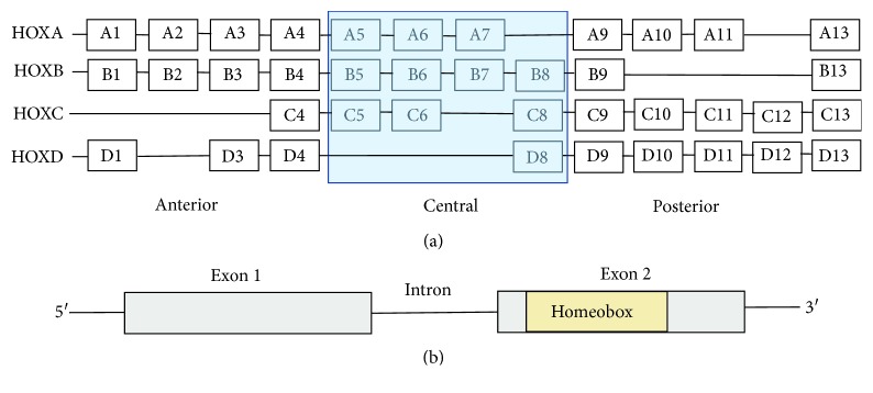
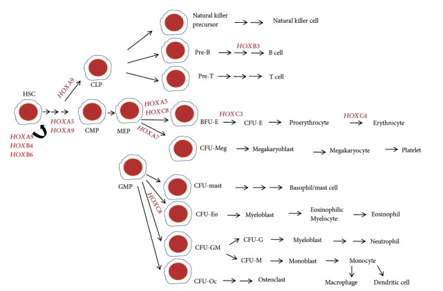
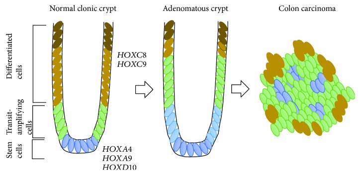
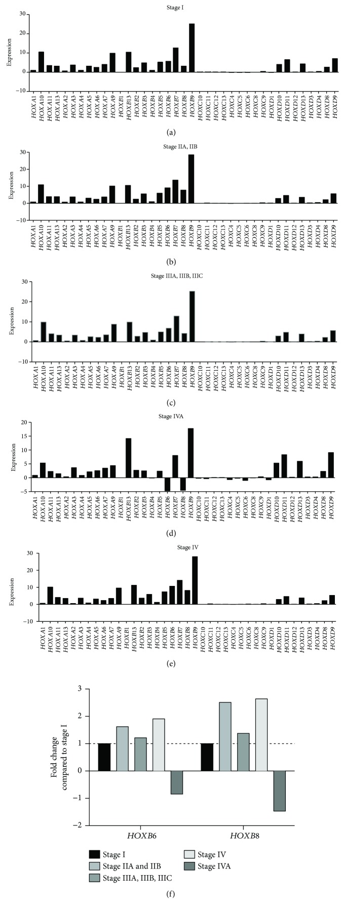
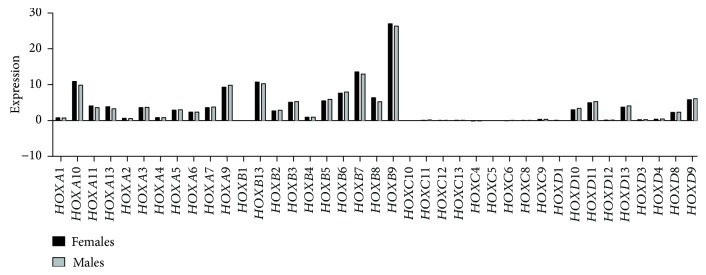
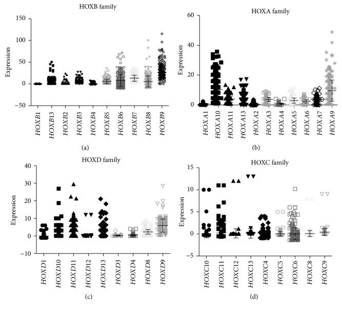
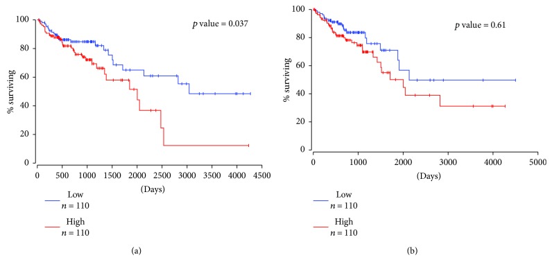
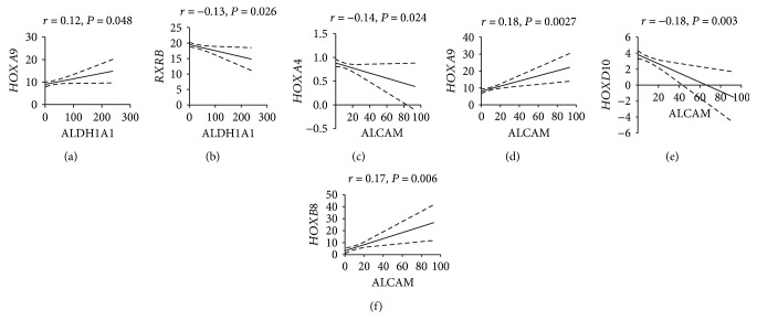
HOX genes encode an evolutionarily conserved set of transcription factors that control how the phenotype of an organism becomes organized during development based on its genetic makeup. For example, in bilaterian-type animals, HOX genes are organized in gene clusters that encode anatomic segment identity, that is, whether the embryo will form with bilateral symmetry with a head (anterior), tail (posterior), back (dorsal), and belly (ventral). Although HOX genes are known to regulate stem cell (SC) differentiation and HOX genes are dysregulated in cancer, the mechanisms by which dysregulation of HOX genes in SCs causes cancer development is not fully understood. Therefore, the purpose of this manuscript was (i) to review the role of HOX genes in SC differentiation, particularly in embryonic, adult tissue-specific, and induced pluripotent SC, and (ii) to investigate how dysregulated HOX genes in SCs are responsible for the development of colorectal cancer (CRC) and acute myeloid leukemia (AML). We analyzed HOX gene expression in CRC and AML using information from The Cancer Genome Atlas study. Finally, we reviewed the literature on HOX genes and related therapeutics that might help us understand ways to develop SC-specific therapies that target aberrant HOX gene expression that contributes to cancer development.


Figures

Figure 1
Figure 1
HOX genes and genome organization. (a) In humans, there are a total of 39, clustered into four families, namely, HOXA, HOXB, HOXC, and HOXD. Each family consists of 13 paralogous groups with nine to eleven numbers assigned based on their sequence similarity and position within the cluster. (b) HOX genes have two exons and 1 intron. Exon 2 has a 120-nucleotide sequence, called a Homeobox that encodes a 61 amino acid HOX protein.
Figure 2
Figure 2
HOX gene expression during hematopoiesis. The hematopoietic stem cell (HSC) is a multipotent stem cell that has the ability to give rise to common lymphoid progenitor (CLP) and common myeloid progenitor (CMP) cells. HOXA9, HOXB4, and HOXB6 are known to be expressed in HSC and regulate HSC self-renewal. HOXA5 and HOXA9 are involved in the proliferation and differentiation of HSC to CMP, and HOXA9 regulates the differentiation of HSC into CLP. HOXB3 is expressed during the differentiation of pre-B cells into B cells. HOXA5 and HOXC8 are expressed during erythroid differentiation of megakaryocyte-erythrocyte progenitors (MEP) whereas HOXA7 is expressed during megakaryocyte differentiation. HOXC3 and HOXC4 are crucial during erythroid lineage differentiation. HOXC8 is shown to play a regulatory role during the differentiation of granulocyte-monocyte progenitor (GMP) cells. HSC: hematopoietic stem cells; CMP: common myeloid progenitor; CLP: common lymphoid progenitor; MEP: megakaryocyte-erythrocyte progenitor; GMP: granulocyte-monocyte progenitor; BFU-E: erythroid burst-forming units; CFU-E: erythroid colony-forming unit; CFU-Meg: megakaryocyte colony-forming unit; CFU-mast: mast colony-forming unit; CFU-Eo: eosinophil colony-forming unit; CFU-GM: granulocyte-monocyte colony-forming unit; CFU-Oc: osteoclasts colony-forming unit.
Figure 3
Figure 3
HOX gene expression during colonocyte differentiation. Normal colonic crypts consist of mainly three types of cells based on their location in the crypt. Colon stem cells (SCs) reside at the base of the colonic crypt (shown in blue color). HOXA4, HOXA9, and HOXD10 are expressed in colonic SCs and regulate colonic crypt SC differentiation [49, 50]. SCs generate transit-amplifying cells (shown in green color) that are actively proliferating and differentiating (shown in gold-bronze yellow color) as they move up the axis in the colonic crypt. Finally, fully differentiated or terminally differentiating cells are found at the top of the crypt (shown in brown color). Studies have shown that HOXA family genes are expressed mostly in proliferating colonic cells, and HOXC family genes are expressed in differentiating cells [68]. HOXB and HOXD family genes are expressed throughout the colonic crypts [68]. In colon tumors, the dysregulation of HOXA4 and HOXA9 in colon SCs caused aberrant self-renewal and proliferation, contributing to colon carcinoma [50]. HOXC8 and HOXC9 are expressed in the differentiating cells in the colonic crypt [68].
Figure 4
Figure 4
HOX gene expression during different stages of CRC. RNA sequencing data for CRC patients obtained from The Cancer Genome Atlas (TCGA) for HOX gene expression (normalized FPKM) and analyzed based on different stages of CRC. We studied (a) 55 cases for stage I, (b) 102 cases for stage IIA and IIB combined, (c) 66 cases reporting stage IIIA, IIIB, and IIIC, (d) 39 samples for stage IV, and (e) 4 cases for stage IVA. (f) Fold changes in the expression of HOXB6 and HOXB8 are shown for stages II, III, IV, and IVA compared to stage I.
Figure 5
Figure 5
Gender-based HOX gene expression in CRC. The Cancer Genome Atlas (TCGA) was used to analyze gender-based differences in HOX gene expression in CRC patients. Normalized FPKM expression of HOX genes is plotted for male versus female CRC patients.
Figure 6
Figure 6
HOX family gene expression in CRC. (a) HOXB family gene expression, (b) HOXA family genes, (c) HOXD family genes, and (d) HOXC family genes were analyzed using TCGA RNAseq for n = 273 patient samples. y-axis denotes normalized FPKM values for HOX gene expression.
Figure 7
Figure 7
Overall survival analysis for HOXA4 and HOXD10 in colorectal cancer. Kaplan-Meier survival analysis of the 220 colorectal cancer patients using TCGA dataset. (a) HOXD10 and (b) HOXA4 survival analysis was performed for CRC patients with a cutoff value of 25th percentile. Credits: http://www.oncolnc.org.
Figure 8
Figure 8
Correlation analysis of HOX genes and retinoid receptors with stem cell markers in colorectal cancer. (a) The expression of HOXA9 and ALDH1A1 (SC marker) in colorectal cancer (CRC) patients is correlated by Pearson correlation. A positive significant correlation was observed between HOXA9 and ALDH1A1 (r = 0.12, P = 0.048). (b) The expression of retinoid receptor RXRB and ALDH1A1 in CRC patients is correlated by Pearson correlation. A negative significant correlation was observed between RXRB and ALDH1A1 (r = −0.13, P = 0.026). (c) The expression of HOXA4 and ALCAM (CD166, SC marker) in CRC patients correlated by Pearson correlation. A negative significant correlation was observed between HOXA4 and ALCAM (r = −0.14, P = 0.024). (d) The expression of HOXA9 and ALCAM in CRC patients correlated by Pearson correlation. A positive significant correlation was observed between HOXA9 and ALCAM (r = 0.18, P = 0.0027). (e) The expression of HOXD10 and ALCAM in CRC patients correlated by Pearson correlation. A negative significant correlation was observed between HOXD10 and ALCAM (r = −0.18, P = 0.003). (f) The expression of HOXB8 and ALCAM in CRC patients correlated by Pearson correlation. A positive significant correlation was observed between HOXB8 and ALCAM (r = −0.17, P = 0.006).
Figure 9
Figure 9
Overall survival analysis for HOXA9 in acute myeloid leukemia. Kaplan-Meier survival analysis of HOXA9 in acute myeloid leukemia (AML) using TCGA dataset. Survival analysis was performed for AML patients (total n = 74) with a cutoff value of 25th percentile. Credits: http://www.oncolnc.org.

References

    1. Quinonez S. C., Innis J. W. Human HOX gene disorders. Molecular Genetics and Metabolism. 2014;111(1):4–15. doi: 10.1016/j.ymgme.2013.10.012. - DOI - PubMed
    1. Loring J. F., Porter J. G., Seilhammer J., Kaser M. R., Wesselschmidt R. A gene expression profile of embryonic stem cells and embryonic stem cell-derived neurons. Restorative Neurology and Neuroscience. 2001;18(2-3):81–88. - PubMed
    1. Deschamps J., van Nes J. Developmental regulation of the Hox genes during axial morphogenesis in the mouse. Development. 2005;132(13):2931–2942. doi: 10.1242/dev.01897. - DOI - PubMed
    1. Maconochie M., Nonchev S., Morrison A., Krumlauf R. Paralogous Hox genes:function and regulation. Annual Review of Genetics. 1996;30(1):529–556. doi: 10.1146/annurev.genet.30.1.529. - DOI - PubMed
    1. Dupé V., Davenne M., Brocard J., et al. In vivo functional analysis of the Hoxa-1 3′ retinoic acid response element (3'RARE) Development. 1997;124(2):399–410. - PubMed

LinkOut - more resources