. 2018 Jul 30:2018:6852303.
doi: 10.1155/2018/6852303. eCollection 2018.

The Effect of 40-Hz Light Therapy on Amyloid Load in Patients with Prodromal and Clinical Alzheimer's Disease

Affiliations

The Effect of 40-Hz Light Therapy on Amyloid Load in Patients with Prodromal and Clinical Alzheimer's Disease

Rola Ismail et al. Int J Alzheimers Dis. .

Abstract

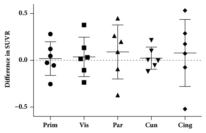
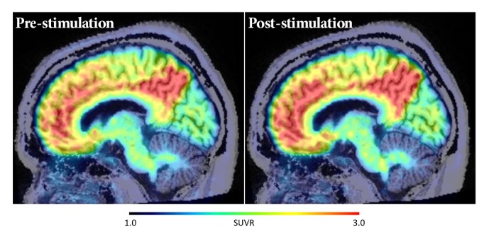
Alzheimer's disease (AD) is a progressive neurodegenerative disorder. AD pathology is characterized by abnormal aggregation of the proteins amyloid-β (Aβ) and hyperphosphorylated tau. No effective disease modifying therapies are currently available. A short-duration intervention with 40 Hz light flicker has been shown to reduce brain Aβ load in transgenic mice. We aimed to test the effect of a similar short-duration 40 Hz light flicker regime in human AD patients. We utilized a Light Emitting Diode (LED) light bulb with a 40 Hz flicker. Six Aβ positive patients received 10 days of light therapy, had 2 hours of daily exposure, and underwent a postintervention PiB PET on day 11. After 10 days of light therapy, no significant decrease of PiB SUVR values was detected in any volumes of interest tested (primary visual cortex, visual association cortex, lateral parietal cortex, precuneus, and posterior cingulate) or in the total motor cortex, and longer treatments may be necessary to induce amyloid removal in humans.


Figures

Figure 1
Figure 1
Mean PiB SUVR differences in five cortical regions. No significant difference in SUVR before and after light stimulation was seen (Prim = primary visual, Vis = visual association, Par = Parietal lobe, Cun = Cuneus, and Cing = posterior cingulate).
Figure 2
Figure 2
Average 11C-PiB SUVR images of the six subjects in the pre- and poststimulation conditions. No significant differences were seen anywhere in cortical regions.

References

    1. Parbo P., Ismail R., Hansen K. V., et al. Brain inflammation accompanies amyloid in the majority of mild cognitive impairment cases due to Alzheimer's disease. Brain. 2017;140(7):2002–2011. doi: 10.1093/brain/awx120. - DOI - PubMed
    1. Braak H., Braak E. Neuropathological stageing of Alzheimer-related changes. Acta Neuropathologica. 1991;82(4):239–259. doi: 10.1007/BF00308809. - DOI - PubMed
    1. Wiley C. Neuropathology and pathogenesis of encephalitis following amyloid-β immunization in alzheimer's disease. Yearbook of Pathology and Laboratory Medicine. 2006;2006:186–188. doi: 10.1016/S1077-9108(08)70140-3. - DOI - PubMed
    1. Sevigny J., Chiao P., Bussière T., et al. The antibody aducanumab reduces Aβ plaques in Alzheimer’s disease. Nature. 2016;537(7618):50–56. doi: 10.1038/nature19323. - DOI - PubMed
    1. Iaccarino H. F., Singer A. C., Martorell A. J., et al. Gamma frequency entrainment attenuates amyloid load and modifies microglia. Nature. 2016;540(7632):230–235. doi: 10.1038/nature20587. - DOI - PMC - PubMed