Cytotoxicity evaluation of chlorhexidine gluconate on human fibroblasts, myoblasts, and osteoblasts
- PMID: 30155401
- PMCID: PMC6098817
- DOI: 10.7150/jbji.26355
Cytotoxicity evaluation of chlorhexidine gluconate on human fibroblasts, myoblasts, and osteoblasts
Abstract
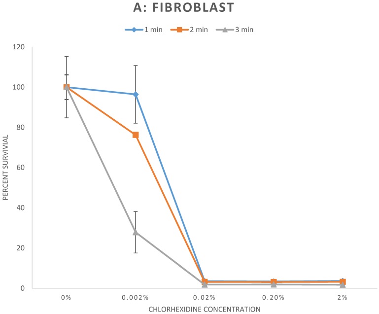
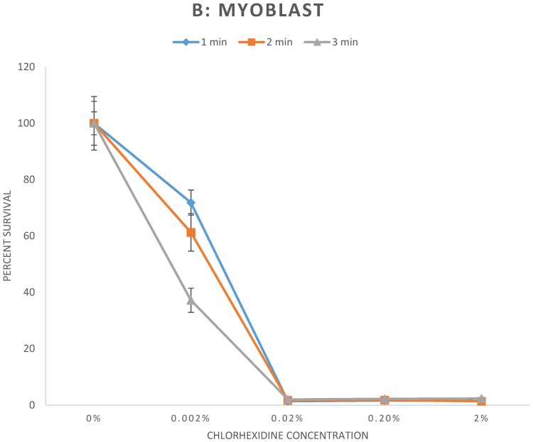
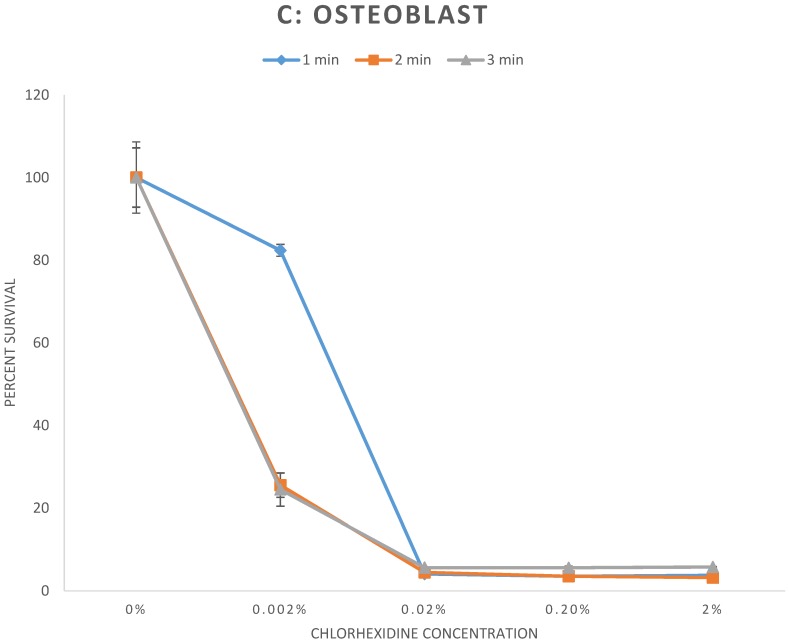
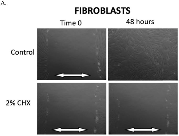
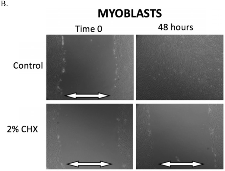
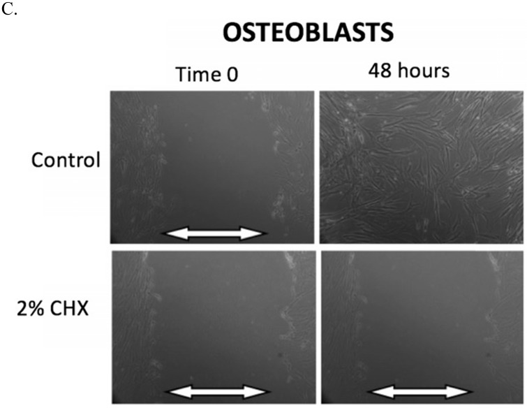
Introduction: Chlorhexidine gluconate (CHX) is widely used as a preoperative surgical skin-preparation solution and intra-wound irrigation agent, with excellent efficacy against wide variety of bacteria. The cytotoxic effect of CHX on local proliferating cells following orthopaedic procedures is largely undescribed. Our aim was to investigate the in vitro effects of CHX on primary fibroblasts, myoblasts, and osteoblasts. Methods: Cells were exposed to CHX dilutions (0%, 0.002%, 0.02%, 0.2%, and 2%) for either a 1, 2, or 3-minute duration. Cell survival was measured using a cytotoxicity assay (Cell Counting Kit-8). Cell migration was measured using a scratch assay: a "scratch" was made in a cell monolayer following CHX exposure, and time to closure of the scratch was measured. Results: All cells exposed to CHX dilutions of ≥ 0.02% for any exposure duration had cell survival rates of less than 6% relative to untreated controls (p < 0.001). Cells exposed to CHX dilution of 0.002% all had significantly lower survival rates relative to control (p < 0.01) with the exception of 1-minute exposure to fibroblasts, which showed 96.4% cell survival (p = 0.78). Scratch defect closure was seen in < 24 hours in all control conditions. However, cells exposed to CHX dilutions ≥ 0.02% had scratch defects that remained open indefinitely. Conclusions: The clinically used concentration of CHX (2%) permanently halts cell migration and significantly reduces survival of in vitro fibroblasts, myoblasts, and osteoblasts. Further in vivo studies are required to examine and optimize CHX safety and efficacy when applied near open incisions or intra-wound application.
Keywords: chlorhexidine; cytotoxicity; fibroblasts; myoblasts; osteoblasts.
Conflict of interest statement
Competing Interests: The authors have declared that no competing interest exists.
Figures
Similar articles
-
Povidone-iodine Solutions Inhibit Cell Migration and Survival of Osteoblasts, Fibroblasts, and Myoblasts.Spine (Phila Pa 1976). 2017 Dec 1;42(23):1757-1762. doi: 10.1097/BRS.0000000000002224. Spine (Phila Pa 1976). 2017. PMID: 28505031
-
Effect of essential oil and chlorhexidine mouthwashes on gingival fibroblast survival and migration.J Periodontol. 2013 Aug;84(8):1211-20. doi: 10.1902/jop.2012.120312. Epub 2012 Oct 29. J Periodontol. 2013. PMID: 23106509
-
Combined and independent cytotoxicity of sodium hypochlorite, ethylenediaminetetraacetic acid and chlorhexidine.Int Endod J. 2016 Aug;49(8):764-73. doi: 10.1111/iej.12517. Epub 2015 Sep 1. Int Endod J. 2016. PMID: 26242704
-
Chlorhexidine gluconate.Aust Endod J. 2005 Aug;31(2):48-52. doi: 10.1111/j.1747-4477.2005.tb00221.x. Aust Endod J. 2005. PMID: 16128251 Review.
-
Efficacy of chlorhexidine rinses after periodontal or implant surgery: a systematic review.Clin Oral Investig. 2019 Jan;23(1):21-32. doi: 10.1007/s00784-018-2761-y. Epub 2018 Dec 7. Clin Oral Investig. 2019. PMID: 30535817
Cited by
-
An in vitro comparison of antimicrobial efficacy and cytotoxicity between povidone-iodine and chlorhexidine for treating clinical endometritis in dairy cows.PLoS One. 2022 Jul 8;17(7):e0271274. doi: 10.1371/journal.pone.0271274. eCollection 2022. PLoS One. 2022. PMID: 35802692 Free PMC article.
-
Development of Solid Nanosystem for Delivery of Chlorhexidine with Increased Antimicrobial Activity and Decreased Cytotoxicity: Characterization and In Vitro and In Ovo Toxicological Screening.Molecules. 2025 Jan 3;30(1):162. doi: 10.3390/molecules30010162. Molecules. 2025. PMID: 39795218 Free PMC article.
-
In-vitro antibiofilm activity of chlorhexidine digluconate on polylactide-based and collagen-based membranes.BMC Oral Health. 2019 Dec 26;19(1):291. doi: 10.1186/s12903-019-0979-y. BMC Oral Health. 2019. PMID: 31878907 Free PMC article.
-
Clinical use and applications of a citrate-based antiseptic lavage for the prevention and treatment of PJI.Front Med (Lausanne). 2024 Jul 2;11:1397192. doi: 10.3389/fmed.2024.1397192. eCollection 2024. Front Med (Lausanne). 2024. PMID: 39015785 Free PMC article.
-
Development of Biocomposite Polymeric Systems Loaded with Antibacterial Nanoparticles for the Coating of Polypropylene Biomaterials.Polymers (Basel). 2020 Aug 15;12(8):1829. doi: 10.3390/polym12081829. Polymers (Basel). 2020. PMID: 32824142 Free PMC article.
References
-
- Barker FG 2nd. Efficacy of prophylactic antibiotic therapy in spinal surgery: a meta-analysis. Neurosurgery. 2002;51(2):391–400. discussion 400-391. - PubMed
-
- Rubinstein E, Findler G, Amit P, Shaked I. Perioperative prophylactic cephazolin in spinal surgery. A double-blind placebo-controlled trial. J Bone Joint Surg Br. 1994;76(1):99–102. - PubMed
-
- Chiang HY, Herwaldt LA, Blevins AE, Cho E, Schweizer ML. Effectiveness of local vancomycin powder to decrease surgical site infections: a meta-analysis. Spine J. 2014;14(3):397–407. - PubMed
-
- van Meurs SJ, Gawlitta D, Heemstra KA, Poolman RW, Vogely HC, Kruyt MC. Selection of an optimal antiseptic solution for intraoperative irrigation: an in vitro study. J Bone Joint Surg Am. 2014;96(4):285–291. - PubMed
LinkOut - more resources
Full Text Sources
Other Literature Sources