

Exploring the Microbiota of Diabetic Foot Infections With Culturomics
- PMID: 30155447
- PMCID: PMC6102383
- DOI: 10.3389/fcimb.2018.00282
Exploring the Microbiota of Diabetic Foot Infections With Culturomics
Abstract
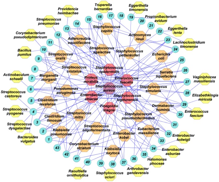
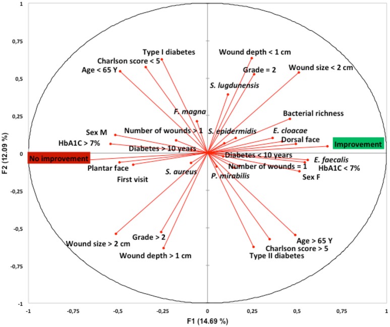
The purpose of this prospective observational study was to evaluate the richness and diversity of bacteria in samples from diabetic foot infections using a culturomics approach. Bacterial culture findings from wound samples were analyzed together with clinical characteristics and treatment outcomes. We included 43 patients admitted to a French referral center with a moderate to severe diabetic foot infection. The 30,000 colonies identified yielded 53 different bacterial species. The global α-Shannon diversity was 3.34 and the bacterial richness per patient was 4 ± 2. Of all the identified bacterial species, 19 (35.8%) had never been previously cultured or identified by molecular methods from diabetic foot ulcers. Most of the samples were polymicrobial (N = 38; 88.3%). Of all the isolated species, the most prevalent were Staphylococcus aureus (N = 28; 52.8%), Enterococcus faecalis (N = 24; 45.2%), Enterobacter cloacae (N = 12; 22.6%), Staphylococcus lugdunensis (N = 10; 18.7%), Staphylococcus epidermidis (N = 6; 11.3%), Proteus mirabilis (N = 6; 11.3%), and Finegoldia magna (N = 5; 9.4%). The only factor associated with wound improvement after a 1-month follow-up was the presence of E. faecalis (p = 0.012) when compared with patients without wound improvement. This study confirms the complementary role of culturomics in the exploration of complex microbiota. Further studies on a larger scale are needed to fully understand the clinical importance of the microbiota of diabetic foot infections.
Keywords: bacterial species; culturomics; diabetes; foot infection; microbiota.
Figures
References
-
- Claros M., Citron D. M., Goldstein E. J., Merriam C. V., Tyrrell K. L. (2013). Differences in distribution and antimicrobial susceptibility of anaerobes isolated from complicated intra-abdominal infections versus diabetic foot infections. Diagn. Microbiol. Infect. Dis. 76, 546–548. 10.1016/j.diagmicrobio.2013.04.025 - DOI - PubMed
Publication types
MeSH terms
LinkOut - more resources
Full Text Sources
Other Literature Sources
Medical
Molecular Biology Databases