. 2018 Aug 2:2018:5647981.
doi: 10.1155/2018/5647981. eCollection 2018.

Markers of Local Inflammation and Bone Resorption in the Acute Diabetic Charcot Foot

Affiliations

Markers of Local Inflammation and Bone Resorption in the Acute Diabetic Charcot Foot

Rasmus Bo Jansen et al. J Diabetes Res. .

Abstract

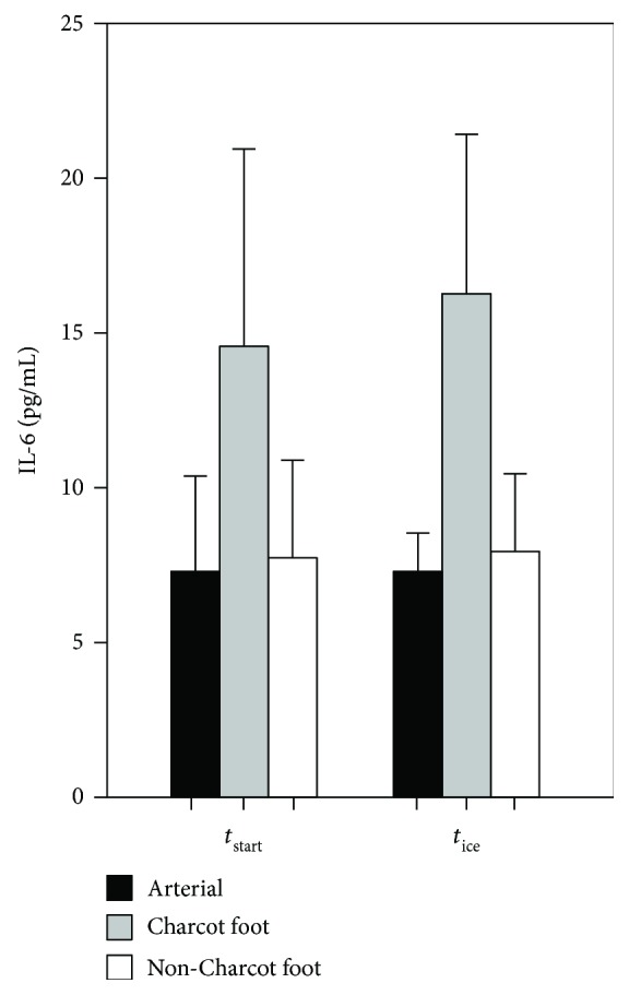
Objective: Due to the localized nature of Charcot foot, systemically altered levels of inflammation markers can be difficult to measure. The aim of this study was to investigate whether it is possible to detect an arteriovenous (A-V) flux in any locally produced inflammatory biomarkers from an acute Charcot foot by comparing local and systemic measurements.

Methods: We included patients with acute diabetic Charcot foot. Blood was sampled from the vena saphena magna on the distal part of the crus bilaterally as well as from the arteria radialis. To minimize the A-V shunting effect, the feet were externally cooled with ice water prior to resampling.

Results: Both before and after cooling, the A-V flux of interleukin-6 (IL-6) between the Charcot feet and the arterial level was significantly higher than the flux between the healthy feet and the arterial level (Δvaluebefore: 7.25 versus 0.41 pg/mL, resp., p = 0.008; Δvalueafter: 10.04 versus 1.68 pg/mL, resp., p = 0.032). There were no differences in the fluxes for other markers of inflammation.

Conclusion: We have found an increased A-V flux of IL-6 in the acute diabetic Charcot foot compared to the healthy foot in the same patients.


Figures

Figure 1
Figure 1
Sites of arterial (red) or venous (blue) blood samples before (t start) and after (t ice) external cooling of the feet. Arterial samples were taken from the a. radialis, or from the a. brachialis if the a. radialis was inaccessible. Venous samples were taken from a large superficial vein at the third distal part of the crus both on the side with a Charcot foot (CF(v)) and on the side without a Charcot foot (non-CF(v)).
Figure 2
Figure 2
Levels of interleukin 6 (IL-6) in arterial and local venous samples in both feet (CF(v) and non-CF(v)) before (t start) and after (t ice) external cooling with ice water. Bars = mean; error bars = SEM.

Similar articles

Cited by

References

    1. Hartemann-Heurtier A., van G. H., Grimaldi A. The Charcot foot. The Lancet. 2002;360(9347):1776–1779. doi: 10.1016/S0140-6736(02)11671-0. - DOI - PubMed
    1. Fabrin J., Larsen K., Holstein P. E. Long-term follow-up in diabetic Charcot feet with spontaneous onset. Diabetes Care. 2000;23(6):796–800. doi: 10.2337/diacare.23.6.796. - DOI - PubMed
    1. Lee L., Blume P. A., Sumpio B. Charcot joint disease in diabetes mellitus. Annals of Vascular Surgery. 2003;17(5):571–580. doi: 10.1007/s10016-003-0039-5. - DOI - PubMed
    1. Rogers L. C., Frykberg R. G. The Charcot foot. The Medical Clinics of North America. 2013;97(5):847–856. doi: 10.1016/j.mcna.2013.04.003. - DOI - PubMed
    1. Grear B. J., Rabinovich A., Brodsky J. W. Charcot arthropathy of the foot and ankle associated with rheumatoid arthritis. Foot & Ankle International. 2013;34(11):1541–1547. doi: 10.1177/1071100713500490. - DOI - PubMed