

Smaller subcortical volume in Parkinson patients with rapid eye movement sleep behavior disorder
- PMID: 30155787
- PMCID: PMC6395547
- DOI: 10.1007/s11682-018-9939-4
Smaller subcortical volume in Parkinson patients with rapid eye movement sleep behavior disorder
Abstract
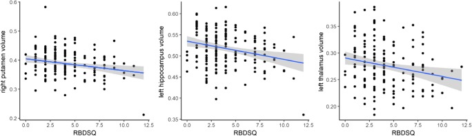
Parkinson disease (PD) patients with rapid eye movement (REM) sleep behavior disorder (RBD) have worse motor symptoms and non-motor symptoms than patients without RBD. The aim of this study was to examine underlying differences in brain structure from a network perspective. Baseline data were obtained from Parkinson's Progression Markers Initiative (PPMI) participants. We divided PD patients and healthy controls (HC) into RBD positive and RBD negative using a cutoff score of ≥5 on the RBD screening questionnaire. HC with probable RBD were excluded. We first carried out a region-of-interest analysis of structural MRIs using voxel-based morphometry to study volumetric differences for the putamen, thalamus and hippocampus in a cross-sectional design. Additionally, an exploratory whole-brain analysis was performed. To study group differences from a network perspective, we then performed a 'seed-based' analysis of structural covariance, using the bilateral dorsal-caudal putamen, mediodorsal thalamus and anterior hippocampus as seed regions. The volume of the right putamen was smaller in PD patients with RBD. RBD symptom severity correlated negatively with volume of the right putamen, left hippocampus and left thalamus. We did not find any differences in structural covariance between PD patients with and without RBD. Presence of RBD and severity of RBD symptoms in PD are associated with smaller volumes of the putamen, thalamus and hippocampus.
Keywords: Parkinson disease; REM sleep behavior disorder; Structural covariance; Voxel-based morphometry.
Conflict of interest statement
The authors declare that they have no conflict of interest. Odile A. van den Heuvel received a grant from PhotoPharmics, NIH BD2K (U54 EB020403–02, PI: P.M. Thompson) and NIMH (R01 MH113250–01, PI: H.B. Simpson). Ysbrand van der Werf received a grant from PhotoPharmics. Henk W. Berendse received a grant from The Netherlands Organisation for Health Research and Development (ZonMw 836,011,029, PI: E.M.J. Foncke), The Michael J Fox Foundation (Grant ID 12274, PI: A.D. Windhorst), and The Michael J Fox Foundation (Grant ID 12062, PI: A.H. Jacobs). Daniel Weintraub has received research funding or support from Michael J. Fox Foundation for Parkinson’s Research, National Institutes of Health (NINDS), Novartis Pharmaceuticals, Department of Veterans Affairs, Avid Radiopharmaceuticals, Alzheimer’s Disease Cooperative Study, and the International Parkinson and Movement Disorder Society; honoraria for consultancy from Acadia, Anavex Life Sciences, Biogen, Biotie (Acorda), BlackThorn Therapeutics, Bracket, Clintrex LLC, Eisai Inc., Eli Lilly, Jazz Pharma, Lundbeck, Roche, Sunovion, Takeda, UCB, and the CHDI Foundation; license fee payments from the University of Pennsylvania for the QUIP and QUIP-RS; royalties from Wolters Kluweland; and fees for legal consultation for three lawsuits related to medication prescribing in patients with Parkinson’s disease. Chris Vriend received a grant from Amsterdam Neuroscience and Brain foundation Netherlands.
Figures
Similar articles
-
Structural Brain Alterations Associated with Rapid Eye Movement Sleep Behavior Disorder in Parkinson's Disease.Sci Rep. 2016 Jun 1;6:26782. doi: 10.1038/srep26782. Sci Rep. 2016. PMID: 27245317 Free PMC article.
-
Reduced thalamic volume in Parkinson disease with REM sleep behavior disorder: volumetric study.Parkinsonism Relat Disord. 2014 Sep;20(9):1004-8. doi: 10.1016/j.parkreldis.2014.06.012. Epub 2014 Jun 24. Parkinsonism Relat Disord. 2014. PMID: 24998995
-
Brain atrophy in Parkinson's disease with polysomnography-confirmed REM sleep behavior disorder.Sleep. 2019 Jun 11;42(6):zsz062. doi: 10.1093/sleep/zsz062. Sleep. 2019. PMID: 30854555 Free PMC article.
-
Brain Neuroimaging of Rapid Eye Movement Sleep Behavior Disorder in Parkinson's Disease: A Systematic Review.J Parkinsons Dis. 2022;12(1):69-83. doi: 10.3233/JPD-212571. J Parkinsons Dis. 2022. PMID: 34806615
-
Interactions of visual hallucinations, rapid eye movement sleep behavior disorder and cognitive impairment in Parkinson's disease: A review.Parkinsonism Relat Disord. 2016 Jan;22:1-8. doi: 10.1016/j.parkreldis.2015.11.018. Epub 2015 Nov 25. Parkinsonism Relat Disord. 2016. PMID: 26639978 Review.
Cited by
-
Abnormal Gray Matter Volume and Functional Connectivity in Parkinson's Disease with Rapid Eye Movement Sleep Behavior Disorder.Parkinsons Dis. 2021 Feb 22;2021:8851027. doi: 10.1155/2021/8851027. eCollection 2021. Parkinsons Dis. 2021. PMID: 33688426 Free PMC article.
-
Neuroimaging Advances in Parkinson's Disease and Atypical Parkinsonian Syndromes.Front Neurol. 2020 Oct 15;11:572976. doi: 10.3389/fneur.2020.572976. eCollection 2020. Front Neurol. 2020. PMID: 33178113 Free PMC article. Review.
-
Cerebellum and basal ganglia connectivity in isolated REM sleep behaviour disorder and Parkinson's disease: an exploratory study.Brain Imaging Behav. 2024 Dec;18(6):1428-1437. doi: 10.1007/s11682-024-00939-x. Epub 2024 Sep 25. Brain Imaging Behav. 2024. PMID: 39320619 Free PMC article.
-
Morphological changes in Parkinson's disease based on magnetic resonance imaging: A mini-review of subcortical structures segmentation and shape analysis.World J Psychiatry. 2022 Dec 19;12(12):1356-1366. doi: 10.5498/wjp.v12.i12.1356. eCollection 2022 Dec 19. World J Psychiatry. 2022. PMID: 36579355 Free PMC article. Review.
-
Disrupted Brain Structural Network Connection in de novo Parkinson's Disease With Rapid Eye Movement Sleep Behavior Disorder.Front Hum Neurosci. 2022 Jul 19;16:902614. doi: 10.3389/fnhum.2022.902614. eCollection 2022. Front Hum Neurosci. 2022. PMID: 35927996 Free PMC article.
References
-
- Arnaldi D, Morbelli S, Brugnolo A et al. (2016) Functional neuroimaging and clinical features of drug naive patients with de novo Parkinson's disease and probable RBD. Parkinsonism & Related Disorders29, 47–53. - PubMed
-
- Behrens, T. E., Johansen-Berg, H., Woolrich, M. W., et al. (2003). Non-invasive mapping of connections between human thalamus and cortex using diffusion imaging. Nature Neuroscience, 6(7), 750–757. - PubMed
-
- Berardelli A, Wenning GK, Antonini A et al. (2013) EFNS/MDS-ES/ENS [corrected] recommendations for the diagnosis of Parkinson's disease. European Journal of Neurology, 20(1), 16–34. - PubMed
-
- Boeve BF. REM sleep behavior disorder: Updated review of the core features, the REM sleep behavior disorder-neurodegenerative disease association, evolving concepts, controversies, and future directions. Annals of the New York Academy of Sciences. 2010;1184:15–54. doi: 10.1111/j.1749-6632.2009.05115.x. - DOI - PMC - PubMed
MeSH terms
Grants and funding
LinkOut - more resources
Full Text Sources
Other Literature Sources
Medical
