Review
. 2018 Sep 26;18(19):2893-2912.
doi: 10.1039/c8lc00330k.

Applications of tumor chip technology

Affiliations
Review

Applications of tumor chip technology

Stephanie J Hachey et al. Lab Chip. .

Abstract

Over the past six decades the inflation-adjusted cost to bring a new drug to market has been increasing constantly and doubles every 9 years - now reaching in excess of $2.5 billion. Overall, the likelihood of FDA approval for a drug (any disease indication) that has entered phase I clinical trials is a mere 9.6%, with the approval rate for oncology far below average at only 5.1%. Lack of efficacy or toxicity is often not revealed until the later stages of clinical trials, despite promising preclinical data. This indicates that the current in vitro systems for drug screening need to be improved for better predictability of in vivo outcomes. Microphysiological systems (MPS), or bioengineered 3D microfluidic tissue and organ constructs that mimic physiological and pathological processes in vitro, can be leveraged across preclinical research and clinical trial stages to transform drug development and clinical management for a range of diseases. Here we review the current state-of-the-art in 3D tissue-engineering models developed for cancer research, with a focus on tumor-on-a-chip, or tumor chip, models. From our viewpoint, tumor chip systems can advance innovative medicine to ameliorate the high failure rates in anti-cancer drug development and clinical treatment.


Conflict of interest statement

7 Conflicts

CCWH is co-founder of Kino Biosciences Inc., a company that develops MPS.

Figures

Fig. 1
Fig. 1. Vascularized micro-tumor (VMT) model
(a) A schematic of the microfluidic platform with a single unit. Three tissue chambers (1 mm × 1 mm × 0.1 mm) constitute 1 unit. Different levels of medium in the four vials drive flow. (b) A schematic of the microfluidic platform with 12 units/plate. (c) GFP+ EC at day 0. (d) A fully-formed vascular network at day 7. (e) A vessel (mCherry, red) wrapped by a pericyte (YFP, yellow) (f) 70 kDa rhodamine dextran flowing through the capillary network (green) formed within the three tissue chambers showing tight barrier function. Tumor cells are labeled in blue. (g) HCT116 colorectal cancer cells (GFP) with vessels (mCherry) are either non-treated (control) or treated with FOLFOX standard chemotherapy on day 7 (0 hour) and imaged every 24 hours. (h) Quantitation of FOLFOX treatment in HCT116 VMT. Tumor significantly regresses with treatment compared to control. Reproduced from Sobrino et al and Phan et al with permission from Nature Publishing Group and the Royal Society of Chemistry.
Fig. 2
Fig. 2. Tumor cell extravasation from in vitro microvessels.
(a) A schematic of a microfluidic device and cell-seeding configuration. Suspended HUVECs form microvascular networks in a gel matrix via paracrine signaling with NHLFs across the central media channel. (b) Photograph of 2-channel microfluidic device. (c) Visualization of VE-cadherin (red) at 60X reveals continuous cell-cell junctions. (d) Collagen IV basement membrane deposition (green) around the lumen (red) and in the perivascular space suggests vessel maturation. (e) Perfusion of vessels with 70 kDa dextran reveals patent lumens void of local leaks. Scale bars are 20 μm. (f) High resolution time-lapse confocal microscopy (40X) of an extravasating entrapped MDA-MB-231 (green). Lumens were labeled with a far-red plasma membrane stain (purple). Tumor cells transmigrate through the endothelium and into the 3D matrix over a period of 4 h. The white arrow at 3:30 h indicates the location of a vessel opening at the site of tumor cell extravasation. Reproduced from Chen et al with permission from the Royal Society of Chemistry.
Fig. 3
Fig. 3. Cancer type-specific modeling on-chip.
(a) (Left) Schematic diagram of a cross-section through 2-channel microfluidic lung-on-a-chip device. (Right) Confocal fluorescence micrograph of a cross-section of the two central cell-lined channels of an alveolus chip. NSCLC cells are labeled with GFP and endothelium with RFP, as shown. Reproduced from Hassell et al with permission from Cell Press, (b) (Left) Workflow for generating bone perivascular (BoPV) niche for studies of breast cancer colonization. (Right Top) Bone tissue reconstruction based on micro-computed tomography (μ-CT) data. (Right Bottom) Rectangular-shaped bone matrix in microfluidic chip. Reproduced from Marturano-Kruik et al with permission from the National Academy of Sciences.
Fig. 4
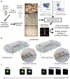
Fig. 4. Examples of onco-immuno chips.
(a) Schematic for preparation and analysis of MDOTS/PDOTS from murine or patient-derived tumor specimens. Reproduced from Jenkins et al with permission from the American Association of Cancer Research. (b) 3D rendering of onco-immuno devices from Pavesi et al. (c) Time-lapse video showing TCR-eT cell killing of HepG2-Env cells on-chip. Reproduced with permission from the American Society for Clinical Investigation.

References

    1. Siegel RL, Miller KD and Jemal A, CA. Cancer J. Clin, 2018, 68, 7–30. - PubMed
    1. Kola I and Landis J, Nat. Rev. Drug Discov, 2004, 3, 711–716. - PubMed
    1. Smietana K, Siatkowski M and Møller M, Nat. Rev. Drug Discov, 2016, 15, 379–380. - PubMed
    1. Waring MJ, Arrowsmith J, Leach AR, Leeson PD, Mandrell S, Owen RM, Pairaudeau G, Pennie WD, Pickett SD,Wang J, Wallace O and Weir A, Nat. Rev. Drug Discov, 2015, 14, 475–486. - PubMed
    1. DiMasi JA, Grabowski HG and Hansen RW, J. Health Econ, 2016, 47, 20–33. - PubMed

Publication types

MeSH terms