

Dermoscopic features of acral melanocytic nevi in a case series from Mexico
- PMID: 30156615
- PMCID: PMC6106682
- DOI: 10.1590/abd1806-4841.20186695
Dermoscopic features of acral melanocytic nevi in a case series from Mexico
Abstract
Background: Pigmented lesions on acral sites are common; clinical differentiation of nevi and early melanoma can be challenging. In these cases, dermoscopy can provide a more accurate diagnosis. Most dermoscopic patterns on acral skin have been described in Asian and European populations, while there are few studies in Latin American populations
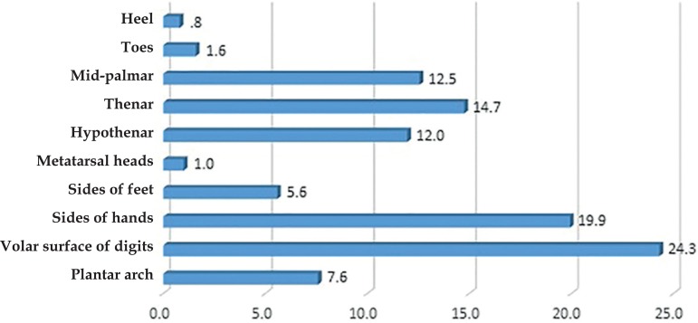
Objectives: To determine the frequency of pigmented lesions in volar skin and their dermoscopic patterns in a Mexican population. Methods: An observational, descriptive, cross-sectional study was performed in Hispanic patients with the presence of at least one pigmented lesion on acral skin. Clinical and dermoscopic images were obtained. These were subsequently evaluated independently by two dermatologists trained and experienced in dermoscopy
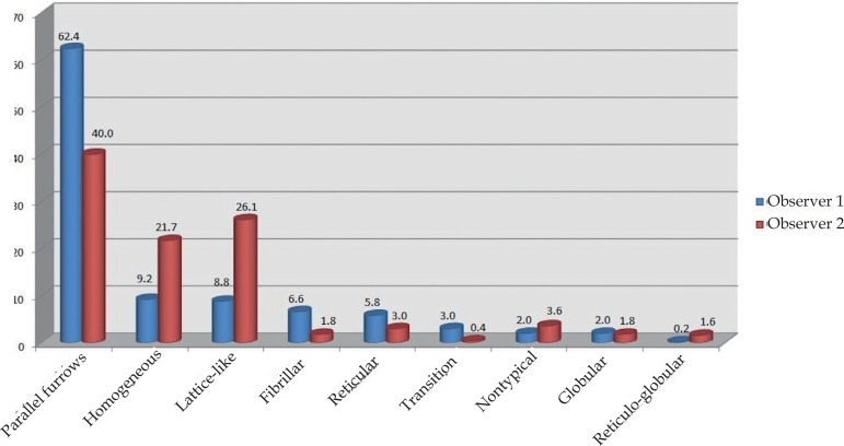
Results: A total of 582 pigmented lesions on volar skin were diagnosed in 321 patients. Overall, prevalence of acral pigmented lesions on volar skin was 6.8%. For both observers, parallel furrows were the most frequent pattern described, but for observer 2, a lattice-like pattern was prevalent on the toes and a homogeneous pattern on the sides of the feet. There was lower inter-observer agreement, with a kappa index of 0.144
Study limitations: The lesions were not biopsied, so clinical-histological correlation could not be performed. The study did not correlate dermoscopic patterns with age
Conclusions: As previously reported by other authors, parallel furrows were the most frequently found dermoscopic pattern on palmoplantar skin
Conflict of interest statement
Conflict of interest: None.
Figures
Similar articles
-
Dermoscopic patterns of 158 acral melanocytic nevi in a Latin American population.Actas Dermosifiliogr. 2013 Sep;104(7):586-92. doi: 10.1016/j.adengl.2013.01.002. Actas Dermosifiliogr. 2013. PMID: 23985085
-
Clinical and Histopathologic Characteristics of Melanocytic Lesions on the Volar Skin Without Typical Dermoscopic Patterns.JAMA Dermatol. 2019 May 1;155(5):578-584. doi: 10.1001/jamadermatol.2018.5926. JAMA Dermatol. 2019. PMID: 30865233 Free PMC article.
-
[Dermoscopic pattern analysis of acral melanocytic nevi].Przegl Lek. 2013;70(11):911-5. Przegl Lek. 2013. PMID: 24697028 Polish.
-
Key points in dermoscopic differentiation between early acral melanoma and acral nevus.J Dermatol. 2011 Jan;38(1):25-34. doi: 10.1111/j.1346-8138.2010.01174.x. J Dermatol. 2011. PMID: 21175752 Review.
-
Dermoscopic patterns of acral melanocytic lesions in skin of color.Cutis. 2019 May;103(5):274-276. Cutis. 2019. PMID: 31233579 Review.
Cited by
-
A European Multicentric Investigation of Atypical Melanocytic Skin Lesions of Palms and Soles: The iDScore-PalmoPlantar Database.Diagnostics (Basel). 2024 Feb 20;14(5):460. doi: 10.3390/diagnostics14050460. Diagnostics (Basel). 2024. PMID: 38472933 Free PMC article.
-
Demystifying the Stinking Reddish Brown Stains Through the Dermoscope: Cydnidae Pigmentation.Indian Dermatol Online J. 2019 Nov 1;10(6):757-758. doi: 10.4103/idoj.IDOJ_346_18. eCollection 2019 Nov-Dec. Indian Dermatol Online J. 2019. PMID: 31807472 Free PMC article. No abstract available.
-
Frequency of Publication of Dermoscopic Images in Inter-observer Studies: A Systematic Review.Acta Derm Venereol. 2021 Dec 17;101(12):adv00621. doi: 10.2340/actadv.v101.865. Acta Derm Venereol. 2021. PMID: 34853864 Free PMC article.
-
Pattern Analysis of Benign and Malignant Atypical Melanocytic Skin Lesions of Palms and Soles: Variations of Dermoscopic Features According to Anatomic Site and Personal Experience.Life (Basel). 2024 May 22;14(6):659. doi: 10.3390/life14060659. Life (Basel). 2024. PMID: 38929643 Free PMC article.
References
-
- Margolis RJ, Tong AK, Byers HR, Mihm Jr MC. Comparison of acral nevomelanocytic proliferations in Japanese and Whites. J Invest Dermatol. 1989;92:222S–226S. - PubMed
-
- Saida T. Malignant melanoma on the sole: how to detect the early lesions efficiently. Pigment Cell Res. 2000;13:135–139. - PubMed
-
- Saida T. Malignant melanoma in situ on the sole of the foot: its clinical and histopathologic characteristics. Am J Dermatopathol. 1989;11:124–130. - PubMed
-
- Saida T, Yoshida N, Ikegawa S, Ishihara K, Nakajima T. Clinical guidelines for the early detection of plantar malignant melanoma. J Am Acad Dermatol. 1990;23:37–40. - PubMed
-
- Tsao H, Atkins MB, Sober AJ. Management of cutaneous melanoma. N Engl J Med. 2004;351:998–1012. - PubMed
Publication types
MeSH terms
LinkOut - more resources
Full Text Sources
Other Literature Sources
Medical
Research Materials
