. 2019 Feb;33(2):1605-1616.
doi: 10.1096/fj.201800965R. Epub 2018 Aug 29.

MicroRNA-570 is a novel regulator of cellular senescence and inflammaging

Affiliations

MicroRNA-570 is a novel regulator of cellular senescence and inflammaging

Jonathan R Baker et al. FASEB J. 2019 Feb.

Abstract

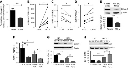
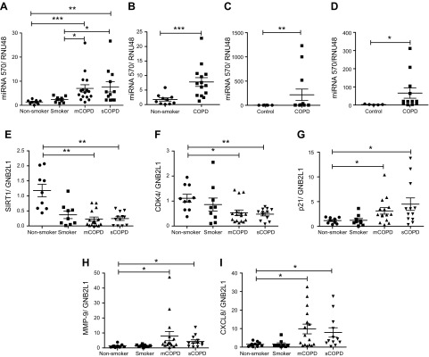
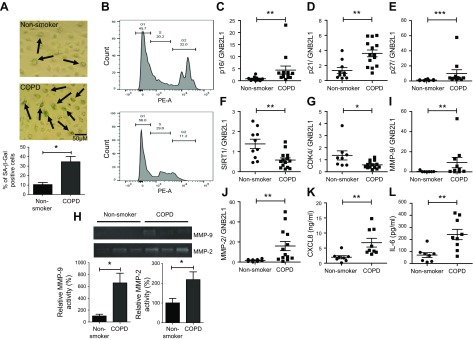
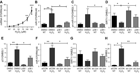
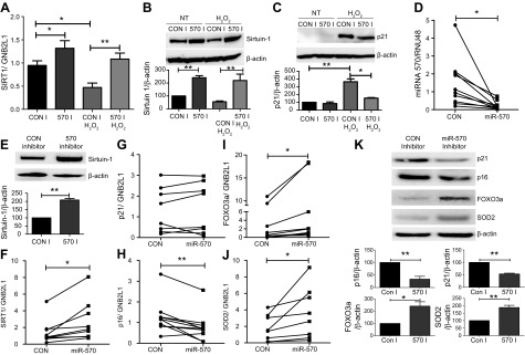
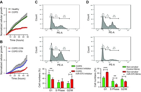
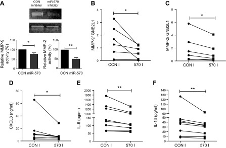
Diseases of accelerated aging often occur together (multimorbidity), and their prevalence is increasing, with high societal and health care costs. Chronic obstructive pulmonary disease (COPD) is one such condition, in which one half of patients exhibit ≥4 age-related diseases. Diseases of accelerated aging share common molecular pathways, which lead to the detrimental accumulation of senescent cells. These senescent cells no longer divide but release multiple inflammatory proteins, known as the senescence-associated secretory phenotype, which may perpetuate and speed disease. Here, we show that inhibiting miR-570-3p, which is increased in COPD cells, reverses cellular senescence by restoring the antiaging molecule sirtuin-1. MiR-570-3p is induced by oxidative stress in airway epithelial cells through p38 MAP kinase-c-Jun signaling and drives senescence by inhibiting sirtuin-1. Inhibition of elevated miR-570-3p in COPD small airway epithelial cells, using an antagomir, restores sirtuin-1 and suppresses markers of cellular senescence (p16INK4a, p21Waf1, and p27Kip1), thereby restoring cellular growth by allowing progression through the cell cycle. MiR-570-3p inhibition also suppresses the senescence-associated secretory phenotype (matrix metalloproteinases-2/9, C-X-C motif chemokine ligand 8, IL-1β, and IL-6). Collectively, these data suggest that inhibiting miR-570-3p rejuvenates cells via restoration of sirtuin-1, reducing many of the abnormalities associated with cellular senescence.-Baker, J. R., Vuppusetty, C., Colley, T., Hassibi, S., Fenwick, P. S., Donnelly, L. E., Ito, K., Barnes, P. J. MicroRNA-570 is a novel regulator of cellular senescence and inflammaging.

Keywords: COPD; cell cycle; epithelial cells; inflammation; miRNA.


Conflict of interest statement

The authors thank Prof. Jim Hogg (University of British Columbia, Vancouver, BC, Canada) for kindly providing peripheral lung tissue samples and Dr. Andriana I. Papaioannou (Sismanogleio Hospital, Athens, Greece) for providing the sputum samples. This work was funded by the British Lung Foundation Grant (JFRG17-7), Wellcome Trust Programme Grant (093080/Z/10/Z), and supported by the NIHR Respiratory Disease Biomedical Research Unit at the Royal Brompton and Harefield NHS Foundation Trust and Imperial College London. The authors declare no conflicts of interest.

Figures

Figure 1
Figure 1
MiRNA-570 directly binds the 3′UTR of sirtuin-1 and is elevated in patients with COPD. A) The dual-luciferase reporter assays using vectors encoding sirtuin-1 target site in the 3′-UTR or control in BEAS-2B cells cotransfected with either an miR-570-3p mimic or mimic control. SAECs from nonsmokers (n = 5) were treated with miR-570-3p mimic or mimic control (CON) for 48 h, miRNA and RNA were extracted, and changes in gene expression assessed by qRT-PCR normalized to guanine nucleotide binding protein-polypeptide 2-like 1 (GNB2L1) or RNU48. B–D) Differences in the gene expression of miRNA-570-3p (B), sirtuin-1 (C), and p21 (D). E) Changes in the protein expression of sirtuin-1 after overexpression of miR-570-3p mimic. BEAS-2B cells were transfected with either an miR-570-3p mimic or mimic control (CON) and treated with or without H2O2 and mRNA or protein extracted. FH) Sirtuin-1 gene expression (F) was assessed, as well as sirtuin-1 (G) and p21 protein (H) (n = 5). Data are means ± sem and were analyzed by using the Mann-Whitney U test, paired or unpaired Student’s t test, or Kruskal-Wallis test with post hoc Dunn’s test; *P < 0.05, **P < 0.01, ***P < 0.001, #P < 0.05.
Figure 2
Figure 2
MiR-570-3p and senescence markers are elevated in lung tissue and cells from patients with COPD. A) Lung tissue from resections obtained from nonsmoker (n = 9) and non-COPD smokers (n = 9), moderate/mild COPD (mCOPD) (n = 16), and severe COPD (sCOPD) (n = 12), and RNA extracted and miR-570-3p expression detected. B) MiR-570-3p levels detected in SAECs from nonsmokers (n = 10) and patients with COPD (n = 14). C) MiR-570-3p levels in peripheral blood mononuclear cells from control [n = 10 (6 nonsmokers and 4 smokers)] and patients with COPD (n = 14). D) MiR-570-3p levels in induced sputum cells from control [n = 5 (1 nonsmoker and 4 smokers)] and patients with COPD (n = 12). E–I) Changes in the gene expression in lung homogenate samples of sirtuin-1 (E), CDK4 (F), p21Waf1 (G), MMP-9 (H), and CXCL8 (I). Data are means ± sem and were analyzed by using a Mann-Whitney U test, paired or unpaired Student’s t test, or Kruskal-Wallis test with post hoc Dunn’s test; *P < 0.05, **P < 0.01, ***P < 0.001.
Figure 3
Figure 3
SAECs from patients with COPD display a cellular senescence phenotype. A) SAECs from nonsmokers and patients with COPD were stained for senescence-associated β-galactosidase (SA-β-Gal), and SA-β-Gal–positive counted (n = 4). B) Untreated passage 2–3 SAECs from nonsmokers and patients with COPD were stained with propidium iodide and fluorescence detected by using flow cytometry on the PE-A channel (n = 4). C–G) p16INK4a (C), p21Waf1 (D), p27Kip1 (E), SIRT1 (F), and CKD4 (G) gene expression detected in SAECs from nonsmokers (n = 8–10) and patients with COPD (n = 11–14). H) Changes in MMP-9 and MMP-2 release from nonsmoker (n = 5) and COPD (n = 5) SAECs were detected by zymography. I, J) MMP-9 (I) and MMP-2 (J) gene expression were also detected in nonsmoker (n = 8–10) and COPD (n = 11–14) SAECs. K, L) Baseline release of CXCL8 (K) and IL-6 (L) from nonsmoker (n = 7) and COPD (n = 8) SAECs, as measured by using ELISA. Data are means ± sem and were analyzed by using a Mann-Whitney U test unpaired Student’s t test, or Kruskal-Wallis test with post hoc Dunn’s test; *P < 0.05, **P < 0.01, ***P < 0.001.
Figure 4
Figure 4
Oxidative stress induces miRNA-570-3p expression in a p38-c-jun–dependent manner. A) MiR-570-3p expression in BEAS-2B cells after H2O2 treatment (n = 5). B–E) p38MAPK inhibitor VX745 (100 nM) effect on miR-570-3p (B), pri-miR-570-3p (C), sirtuin-1 (D), and p21Waf1 (E) gene expression in BEAS-2B cells treated with or without H2O2 (n = 6). F–H) MiR-570-3p (F), sirtuin-1 (G), and p21Waf1 (H) gene expression in BEAS-2B cells were transfected with c-Jun siRNA (100 nM) or random oligonucleotide and then treated with or without H2O2 (n = 6). Data are means ± sem and were analyzed by using Kruskal-Wallis test with post hoc Dunn’s test, 1-way ANOVA with post hoc Bonferroni correction, unpaired or paired Student’s t test, and Wilcoxon signed rank test; *P < 0.05, **P < 0.01, ***P < 0.001.
Figure 5
Figure 5
Inhibition of miR-570-3p rescues sirtuin-1 expression and modulates senescence markers. A–C) BEAS-2B cells (n = 7–10) treated with or without H2O2 and miRNA-570-3p antagomir (60 nM) or control and SIRT1 mRNA (n = 5) (A) and sirtuin-1 (B) and p21Waf1 (C) protein (n = 4) expression were examined. D–F) MiR-570-3p (D), sirtuin-1 protein (E), and SIRT1 mRNA (F) were detected in SAECs from patients with COPD treated with an miR-570-3p antagomir (60 nM) or control (n = 5–10). G–J) In these same cells, p21Waf1 (G), p16INK4a (H), FOXO3a (I), and SOD2 (J) mRNA expression was detected. K) SAECs from patients with COPD treated with an miR-570-3p antagomir (60 nM) or control for 48 h and p21Waf1, p16INK4a, FOXO3a, and SOD2 protein expression were detected (n = 5). Data are means ± sem and were analyzed by using Kruskal-Wallis test with post hoc Dunn’s test, 1-way ANOVA with post hoc Bonferroni correction, unpaired or paired Student’s t test, and Wilcoxon signed rank test; *P < 0.05; **P < 0.01. GNB2L1, guanine nucleotide binding protein-polypeptide 2-like 1.
Figure 6
Figure 6
Inhibition of miRNA-570 expression in airway epithelial cells rescues cellular growth. A) Cellular proliferation measured by using the iCELLigence microelectronic biosensor system of SAECs from nonsmokers and patients with COPD (n = 4). B) Effect of miR-570-3p antagomir or control oligonucleotide on COPD SAECs proliferation (n = 4). C) Effect of miRNA-570-3p antagomir or oligonucleotide control, in COPD SAECs stained with propidium iodide and fluorescence detected by using flow cytometry on the PE-A channel (n = 5). D) Flow cytometric analysis of propidium iodide staining of miRNA-570-3p mimic or oligonucleotide control treated nonsmokers SAECs (n = 4). Data are means ± sem and were analyzed by using 2-way ANOVA with post hoc Bonferroni correction; *P < 0.05, **P < 0.01, ***P < 0.001.
Figure 7
Figure 7
Inhibition of miRNA-570 expression in airway epithelial cells suppresses SASP release. A) Zymography of MMP-2 and MMP-9 expression measured in supernatants from COPD SAECs treated with miRNA-570-3p antagomir or oligonucleotide control (n = 5). B, C) MMP-9 (B) and MMP-2 (C) mRNA expression detected by qRT-PCR in SAECs after treatment with miRNA-570-3p antagomir or oligonucleotide control. D–F) Changes in CXCL8 (D), IL-6 (E), and IL-1β (F) release from COPD SAECs treated with either miRNA-570-3p antagomir or oligonucleotide control (n = 8). Data are means ± sem and were analyzed by using unpaired Student’s t test or Wilcoxon signed rank test; *P < 0.05, **P < 0.01.

References

    1. Lozano, R., Naghavi, M., Foreman, K., Lim, S., Shibuya, K., Aboyans, V., Abraham, J., Adair, T., Aggarwal, R., Ahn, S. Y., Alvarado, M., Anderson, H. R., Anderson, L. M., Andrews, K. G., Atkinson, C., Baddour, L. M., Barker-Collo, S., Bartels, D. H., Bell, M. L., Benjamin, E. J., Bennett, D., Bhalla, K., Bikbov, B., Bin Abdulhak, A., Birbeck, G., Blyth, F., Bolliger, I., Boufous, S., Bucello, C., Burch, M., Burney, P., Carapetis, J., Chen, H., Chou, D., Chugh, S. S., Coffeng, L. E., Colan, S. D., Colquhoun, S., Colson, K. E., Condon, J., Connor, M. D., Cooper, L. T., Corriere, M., Cortinovis, M., de Vaccaro, K. C., Couser, W., Cowie, B. C., Criqui, M. H., Cross, M., Dabhadkar, K. C., Dahodwala, N., De Leo, D., Degenhardt, L., Delossantos, A., Denenberg, J., Des Jarlais, D. C., Dharmaratne, S. D., Dorsey, E. R., Driscoll, T., Duber, H., Ebel, B., Erwin, P. J., Espindola, P., Ezzati, M., Feigin, V., Flaxman, A. D., Forouzanfar, M. H., Fowkes, F. G., Franklin, R., Fransen, M., Freeman, M. K., Gabriel, S. E., Gakidou, E., Gaspari, F., Gillum, R. F., Gonzalez-Medina, D., Halasa, Y. A., Haring, D., Harrison, J. E., Havmoeller, R., Hay, R. J., Hoen, B., Hotez, P. J., Hoy, D., Jacobsen, K. H., James, S. L., Jasrasaria, R., Jayaraman, S., Johns, N., Karthikeyan, G., Kassebaum, N., Keren, A., Khoo, J. P., Knowlton, L. M., Kobusingye, O., Koranteng, A., Krishnamurthi, R., Lipnick, M., Lipshultz, S. E., Ohno, S. L., Mabweijano, J., MacIntyre, M. F., Mallinger, L., March, L., Marks, G. B., Marks, R., Matsumori, A., Matzopoulos, R., Mayosi, B. M., McAnulty, J. H., McDermott, M. M., McGrath, J., Mensah, G. A., Merriman, T. R., Michaud, C., Miller, M., Miller, T. R., Mock, C., Mocumbi, A. O., Mokdad, A. A., Moran, A., Mulholland, K., Nair, M. N., Naldi, L., Narayan, K. M., Nasseri, K., Norman, P., O’Donnell, M., Omer, S. B., Ortblad, K., Osborne, R., Ozgediz, D., Pahari, B., Pandian, J. D., Rivero, A. P., Padilla, R. P., Perez-Ruiz, F., Perico, N., Phillips, D., Pierce, K., Pope C. A. III, Porrini, E., Pourmalek, F., Raju, M., Ranganathan, D., Rehm, J. T., Rein, D. B., Remuzzi, G., Rivara, F. P., Roberts, T., De León, F. R., Rosenfeld, L. C., Rushton, L., Sacco, R. L., Salomon, J. A., Sampson, U., Sanman, E., Schwebel, D. C., Segui-Gomez, M., Shepard, D. S., Singh, D., Singleton, J., Sliwa, K., Smith, E., Steer, A., Taylor, J. A., Thomas, B., Tleyjeh, I. M., Towbin, J. A., Truelsen, T., Undurraga, E. A., Venketasubramanian, N., Vijayakumar, L., Vos, T., Wagner, G. R., Wang, M., Wang, W., Watt, K., Weinstock, M. A., Weintraub, R., Wilkinson, J. D., Woolf, A. D., Wulf, S., Yeh, P. H., Yip, P., Zabetian, A., Zheng, Z. J., Lopez, A. D., Murray, C. J., AlMazroa, M. A., Memish, Z. A. (2012) Global and regional mortality from 235 causes of death for 20 age groups in 1990 and 2010: a systematic analysis for the Global Burden of Disease Study 2010 [published erratum appears in Lancet 2013;381:628] . Lancet 380, 2095–2128 10.1016/S0140-6736(12)61728-0 - DOI - PMC - PubMed
    1. Cosio, M. G., Saetta, M., Agusti, A. (2009) Immunologic aspects of chronic obstructive pulmonary disease. N. Engl. J. Med. 360, 2445–2454 10.1056/NEJMra0804752 - DOI - PubMed
    1. Vanfleteren, L. E., Spruit, M. A., Groenen, M., Gaffron, S., van Empel, V. P., Bruijnzeel, P. L., Rutten, E. P., Op ’t Roodt, J., Wouters, E. F., Franssen, F. M. (2013) Clusters of comorbidities based on validated objective measurements and systemic inflammation in patients with chronic obstructive pulmonary disease. Am. J. Respir. Crit. Care Med. 187, 728–735 10.1164/rccm.201209-1665OC - DOI - PubMed
    1. Barnes, P. J. (2015) Mechanisms of development of multimorbidity in the elderly. Eur. Respir. J. 45, 790–806 10.1183/09031936.00229714 - DOI - PubMed
    1. Kennedy, B. K., Berger, S. L., Brunet, A., Campisi, J., Cuervo, A. M., Epel, E. S., Franceschi, C., Lithgow, G. J., Morimoto, R. I., Pessin, J. E., Rando, T. A., Richardson, A., Schadt, E. E., Wyss-Coray, T., Sierra, F. (2014) Geroscience: linking aging to chronic disease. Cell 159, 709–713 10.1016/j.cell.2014.10.039 - DOI - PMC - PubMed

Publication types

LinkOut - more resources