

Inter-kingdom effect on epithelial cells of the N-Acyl homoserine lactone 3-oxo-C12:2, a major quorum-sensing molecule from gut microbiota
- PMID: 30157234
- PMCID: PMC6114859
- DOI: 10.1371/journal.pone.0202587
Inter-kingdom effect on epithelial cells of the N-Acyl homoserine lactone 3-oxo-C12:2, a major quorum-sensing molecule from gut microbiota
Abstract
Background and aims: N-acyl homoserine lactones (AHLs), which are autoinducer quorum-sensing molecules involved in the bacterial communication network, also interact with eukaryotic cells. Searching for these molecules in the context of inflammatory bowel disease (IBD) is appealing. The aims of our study were to look for AHL molecules in faecal samples from healthy subjects (HS) and IBD patients to correlate AHL profiles with the microbiome and investigate the effect of AHLs of interest on epithelial cells.
Methods: Using mass spectrometry, we characterised AHL profiles in faecal samples from HS (n = 26) and IBD patients in remission (n = 24) and in flare (n = 25) and correlated the presence of AHLs of interest with gut microbiota composition obtained by real-time qPCR and 16S sequencing. We synthesised AHLs of interest to test the inflammatory response after IL1β stimulation and paracellular permeability on Caco-2 cells.
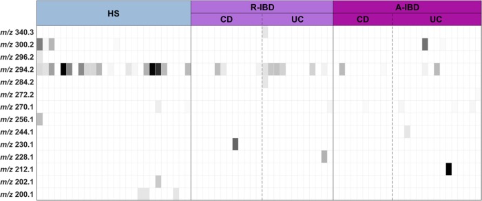
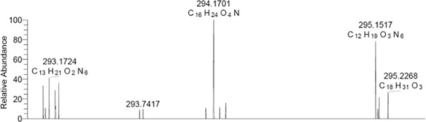
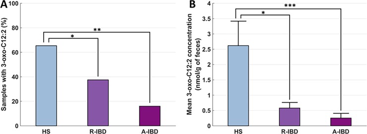
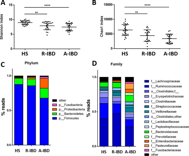
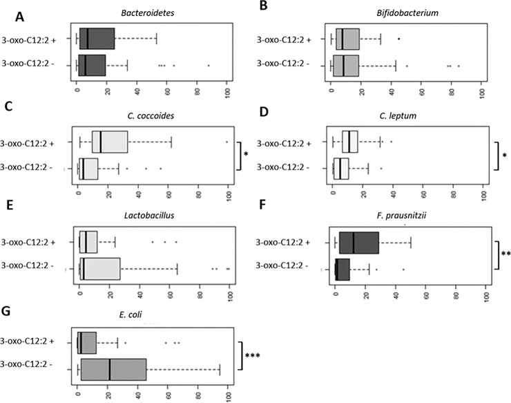
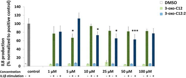
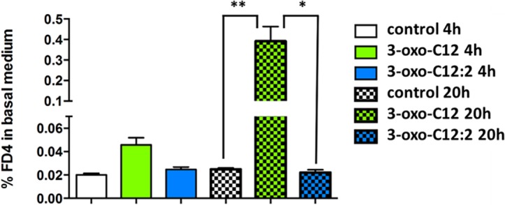
Results: We observed 14 different AHLs, among which one was prominent. This AHL corresponded to 3-oxo-C12:2 and was found significantly less frequently in IBD patients in flare (16%) and in remission (37.5%) versus HS (65.4%) (p = 0.001). The presence of 3-oxo-C12:2 was associated with significantly higher counts of Firmicutes, especially Faecalbacterium prausnitzii, and lower counts of Escherichia coli. In vitro, 3-oxo-C12:2 exerted an anti-inflammatory effect on Caco-2 cells. Interestingly, although 3-oxo-C12, the well-known AHL from Pseudomonas aeruginosa, increased paracellular permeability, 3-oxo-C12:2 did not.
Conclusions: We identified AHLs in the human gut microbiota and discovered a new and prominent AHL, 3-oxo-C12:2, which correlates with normobiosis and exerts a protective effect on gut epithelial cells.
Conflict of interest statement
Dr Landman has received personal fees from Abbvie and Hospira and travel support from Abbvie, Hospira, Mayoly Spindler, Biocodex and Takeda. Professor Marteau has received lecture fees from Abbvie, Astellas, Biocodex, Danone, Hospira-Pfizer, Janssen, Merck-MSD, Ferring Pharmaceuticals and Takeda. Professor Beaugerie has received consulting fees from Janssen; lecture fees from Abbvie, Janssen, MSD, Ferring Pharmaceuticals and Takeda; and research support from Abbvie, Ferring Pharmaceuticals, Hospira-Pfizer, Janssen and Takeda. Professor Sokol has received personal fees from Danone, MSD, Takeda, Abbvie, Astellas, BMS and Novartis; options from Enterome and Maat; and grants from Biocodex. Professor Seksik has received personal fees from Takeda, Merck MSD, Biocodex and Abbvie and non-financial support from Takeda. The remaining authors disclose no conflict. This does not alter our adherence to PLOS ONE policies on sharing data and materials.
Figures

References
-
- Sokol H, Pigneur B, Watterlot L, Lakhdari O, Bermúdez-Humarán LG, Gratadoux J-J, et al. Faecalibacterium prausnitzii is an anti-inflammatory commensal bacterium identified by gut microbiota analysis of Crohn disease patients. Proc Natl Acad Sci. 2008;105: 16731–16736. 10.1073/pnas.0804812105 - DOI - PMC - PubMed
Publication types
MeSH terms
Substances
LinkOut - more resources
Full Text Sources
Other Literature Sources
