. 2018 Aug 29;13(8):e0203135.
doi: 10.1371/journal.pone.0203135. eCollection 2018.

Selective ablation of striatal striosomes produces the deregulation of dopamine nigrostriatal pathway

Affiliations

Selective ablation of striatal striosomes produces the deregulation of dopamine nigrostriatal pathway

Kirill Shumilov et al. PLoS One. .

Abstract

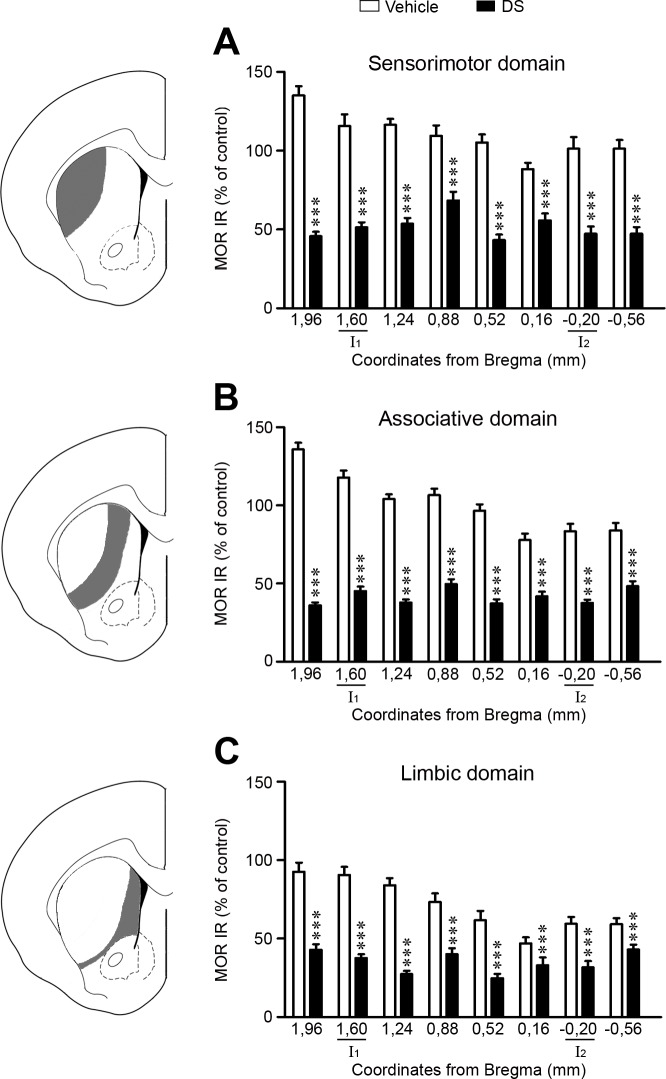
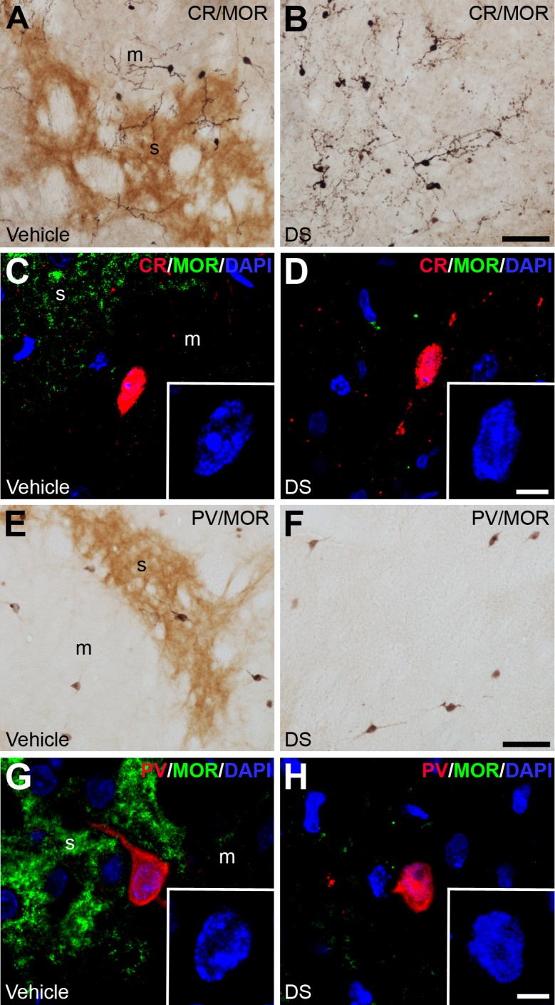
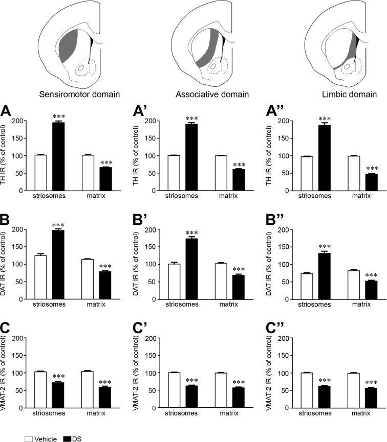
The striatum is a complex structure in which the organization in two compartments (striosomes and matrix) have been defined by their neurochemical profile and their input-output connections. The striosomes receive afferences from the limbic brain areas and send projections to the dopamine neurons of the substantia nigra pars compacta. Thereby, it has been suggested that the striosomes exert a limbic control over the motor function mediated by the surrounding matrix. However, the functionality of the striosomes are not completely understood. To elucidate the role of the striosomes on the regulation of the nigral dopamine neurons, we have induced specific ablation of this compartment by striatal injections of the neurotoxin dermorphin-saporin (DS) and dopamine neurotransmission markers have been analyzed by immunohistochemistry. The degeneration of the striosomes resulted in a nigrostriatal projections imbalance between the two striatal compartments, with an increase of the dopamine neurotransmission in the striosomes and a decrease in the matrix. The present results highlight the key function of the striosomes for the maintenance of the striatal dopamine tone and would contribute to the understanding of their involvement in some neurological disorders such as Huntington's disease.


Conflict of interest statement

The authors have declared that no competing interests exist.

Figures

Fig 1
Fig 1. Schematic representation of procedures.
(A) Diagrams of coronal brain plates modified from a rat brain atlas [26] depicting the sites where the stereotaxic injections were made. (B) Template obtained from a MOR immunolabeled brain section showing the distribution of the striosomes (s) into the dorsal striatum. The template is applied over the photomicrography of a section immunolabeled for TH, DAT or VMAT-2 (in this figure illustrated by TH) to quantify the optical density of each marker in the striosomal (s) and matrix (m) compartments. Abbreviations: AP: anteroposterior; L: lateral; V: ventral. Scale bar is 1 mm.
Fig 2
Fig 2. Unilateral dermorphin-SAP intrastriatal injections produced striosomes ablation.
(A and D) Representative photomicrographs showing the effect of unilateral intrastriatal injections of DS (A) and SAP (D) on MOR IR. Vehicle injected hemisphere was used as control. (B and E) Graphs represent optical density values of MOR IR in the striosomes after DS (#1 to #7; B) and SAP (C1 to C3; E) injection in each rat. Data represent mean ± SEM and are expressed as percentage of vehicle-injected hemisphere. (C and F) Analysis of the number of striosomes in categories ranked by percentages of MOR IR in the rats injected with DS (C) or SAP (F). Data represent mean ± SEM. Statistical analyses of the data were performed with Mann Whitney U test, ***P < 0.001. Scale bars are 1 mm. Abbreviations: Acb: nucleus accumbens; cc: corpus callosum; CPu: caudate putamen; DS: dermorphin-SAP; MOR: μ opioid receptor; SAP: saporin.
Fig 3
Fig 3. Dermorphin-SAP significantly reduced MOR immunoreactivity through the rostro-caudal axis of the caudate putamen.
Graphs represent the optical density values of MOR IR in eight consecutive coronal sections through the rostro-caudal axis of each functional domain of the rat striatum (A: sensorimotor; B: associative; C: limbic). I1 and I2 indicate the anteroposterior Bregma levels of the injection sites. Data represent mean ± SEM and are expressed as percentage of vehicle treated hemisphere. Statistical analysis of the data was performed with Mann Whitney U test, ***P<0.001.
Fig 4
Fig 4. ChAT and SS striatal interneurons are affected by dermorphin-SAP lesion.
(A-C; G-I) Photomicrographs illustrating by dual labeled immunohistochemistry with anti-MOR (brown) and anti-ChAT (A-C) or anti-SS (G-I) (dark blue) the impact of DS on these cells in both the lateral (B-H) and medial part (C-I) of the CPu. (D-F; J-L) Confocal laser photomicrographs illustrating the co-localization of MOR (green) with ChAT (D-F) and SS (J-L) (red). The nuclei are counterstaining with DAPI (blue). Insets show a high magnification of the interneuron nucleus. Abbreviations: CPu: caudate putamen; DS: dermorphin-SAP; m: matrix; s: striosome. Scale bar is 50 μm in A-C, G-I; 10 μm in D-F, J-L; 5 μm in the insets.
Fig 5
Fig 5. CR and PV striatal interneurons are not affected by dermorphin-SAP lesion.
(A-B; E-F) Dual labeled immunohistochemistry with anti-MOR (brown) and anti-CR (A-B) or anti-PV (E-F) (dark blue) to demonstrate the presence of these cells in the vehicle and DS injected hemisphere. (C-D; G-H) Confocal laser photomicrographs illustrating the co-localization of MOR (green) with CR (C-D) and PV (G-H) (red). The nuclei are counterstaining with DAPI (blue). Insets show a high magnification of the interneuron nucleus. Abbreviations: DS: dermorphin-SAP; m: matrix; s: striosome. Scale bar is 50 μm in A-B, E-F; 10 μm in C-D, G-H; 5 μm in the insets.
Fig 6
Fig 6. Striosomal ablation affected nigrostriatal dopamine innervation.
(A-C) Representative photomicrographs illustrating the immunoreactivity of TH (A), MOR (B) and DAT (C) in consecutive sections from rats with unilateral ablation of striosomes. Arrows indicate patches with an enrichment in TH IR and DAT IR in the lesioned hemisphere. (A’-A”, C’-C”) High magnification photomicrographs taken from the areas of the CPu pointed as boxes in (A) and (C) in the unlesioned and lesioned hemispheres. These areas are identified as striosomes in the MOR immunolabeling section (B’-B”). (D-F) Detailed photomicrographs showing TH IR in the CPu of vehicle (D) and DS injected (E and F) hemispheres. An increase in the size of TH IR varicosities in the striosomes (E) and matrix (F) compartments is observed in the DS lesioned CPu. Abbreviations: ac: anterior commissure; Acb: nucleus accumbens; cc: corpus callosum; CPu: caudate putamen; DS: dermorphin-SAP; m, matrix; s, striosome. Scale in C bar is 1 mm (applies to A, B, C), 50 μm in C” (applies to A’-C”) and 50 μm in D (applies to D-F).
Fig 7
Fig 7. Changes in the nigrostriatal dopamine pathway after striosomes ablation.
Graphs represent the semi-quantitative analysis of TH IR (A-A”), DAT IR (B-B”) and VMAT-2 (C-C”) in the striosomes and matrix of vehicle and DS injected hemispheres. The analysis is shown in the three functional domains of the rat CPu (sensorimotor: A, B, C; associative: A’, B’, C’; limbic: A”, B”, C”). Data represent mean ± SEM and are expressed as percentage of vehicle treated hemisphere. Statistical analysis was performed with Student’s t test; *** P < 0.001 DS vs. vehicle.
Fig 8
Fig 8. Striosomal ablation affected a subset of nigral dopamine neurons.
(A) Frequency of nigral cells (expressed as percentage of total cells analyzed) in categories ranked by percentages of TH IR in the control and lesioned hemispheres. Statistical analyses of the data were performed with Student’s t test, *P<0.05, **P<0.01. (B and B’) High magnification photomicrographs showing TH IR cells of the SNc. Scale bar is 5 μm.
Fig 9
Fig 9. Schematic diagram of striosomal circuits into the basal ganglia.
In the striatum the striosomal and matrix compartments are depicted whereas in the SNc the dorsal and ventral tiers are illustrated. Red lines indicate the direct GABAergic projections from the striosomes to the SNc and their collaterals to the GP and SNr. Green lines point the dopaminergic projections from the SNc to the striosomes and matrix. Abbreviations: SNc: substantia nigra pars compacta; SNr: substantia nigra pars reticulata; GP: globus pallidus; STh: subthalamic nucleus; DA: dopamine; Glu: glutamate.

References

    1. Lanciego JL, Luquin N, Obeso JA. Functional neuroanatomy of the basal ganglia. Cold Spring Harb Perspect Med. 2012. December;2(12):a009621–a009621. 10.1101/cshperspect.a009621 - DOI - PMC - PubMed
    1. Balleine BW, Delgado MR, Hikosaka O. The Role of the Dorsal Striatum in Reward and Decision-Making. J Neurosci. Society for Neuroscience; 2007. May 1;27(31):8161–5. - PMC - PubMed
    1. Graybiel AM, Grafton ST. The striatum: Where skills and habits meet. Cold Spring Harb Perspect Biol. 2015;7(8). - PMC - PubMed
    1. Gerfen CR. The neostriatal mosaic: compartmentalization of corticostriatal input and striatonigral output systems. Nature. 1984;311:461–4. - PubMed
    1. Kawaguchi Y, Wilson CJ, Augood SJ, Emson PC. Striatal interneurones: chemical, physiological and morphological characterization. Vol. 18, Trends in Neurosciences. 1995. p. 527–35. - PubMed

Publication types