. 2018 Aug 29;37(1):207.
doi: 10.1186/s13046-018-0878-0.

Astragaloside IV inhibits lung cancer progression and metastasis by modulating macrophage polarization through AMPK signaling

Affiliations

Astragaloside IV inhibits lung cancer progression and metastasis by modulating macrophage polarization through AMPK signaling

Fei Xu et al. J Exp Clin Cancer Res. .

Erratum in

Abstract

Background: Accumulating evidence suggests that M2-polarized tumor-associated macrophages (TAMs) play an important role in cancer progression and metastasis, making M2 polarization of TAMs an ever more appealing target for therapeutic intervention. Astragaloside IV (AS-IV), a saponin component isolated from Astragali radix, has been reported to inhibit the invasion and metastasis of lung cancer, but its effects on TAMs during lung cancer progression have not been investigated.

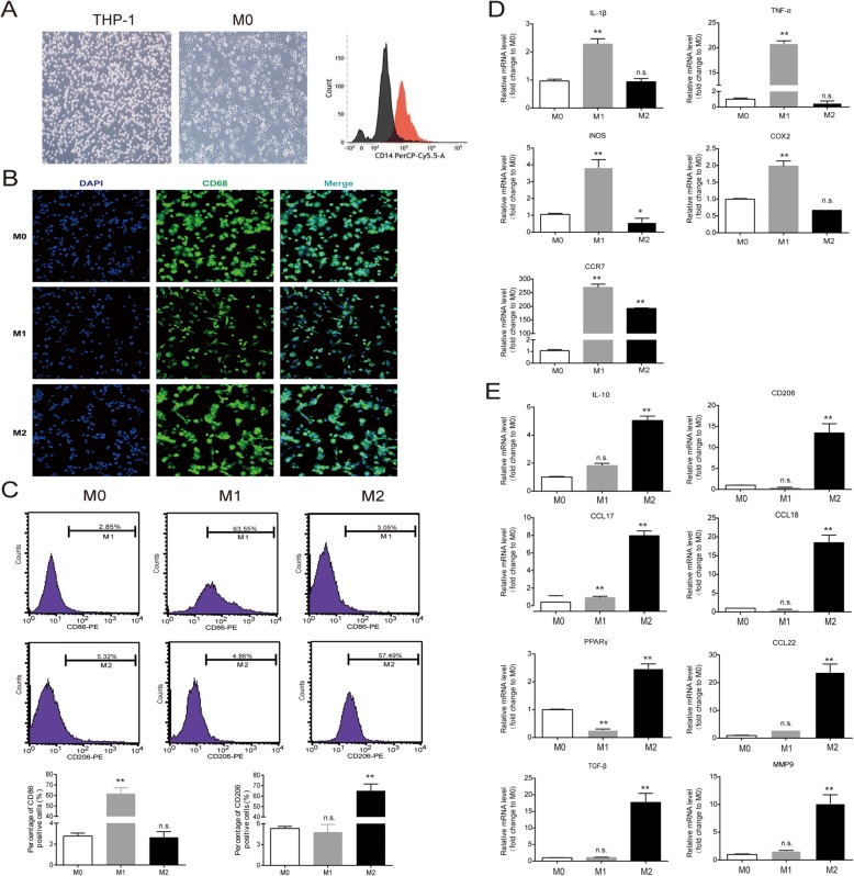
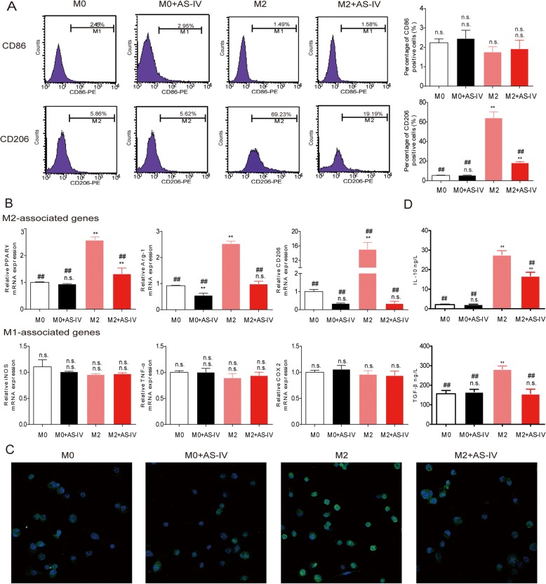
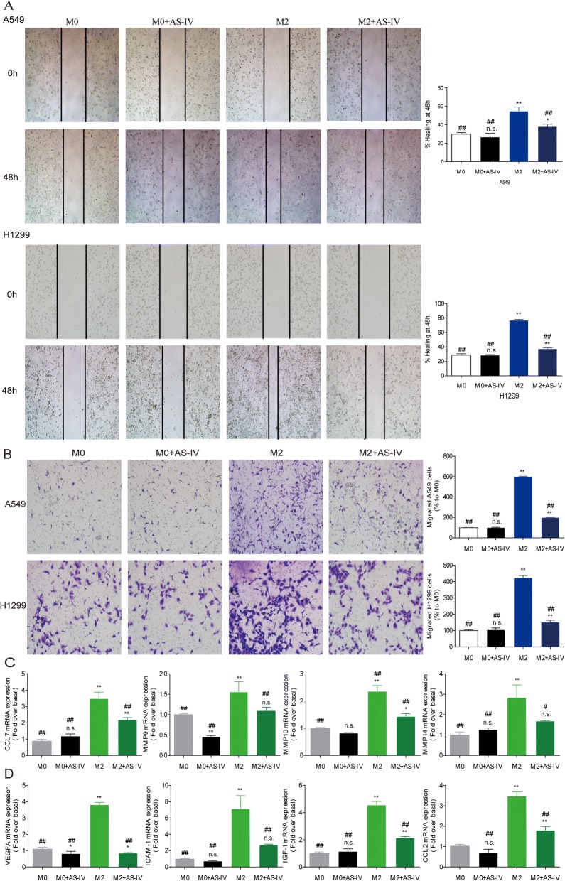
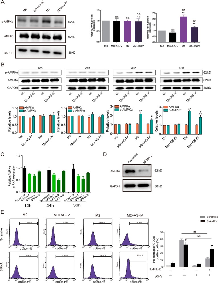
Methods: Human THP-1 monocytes were induced to differentiate into M2 macrophages through treatments with IL-4, IL-13, and phorbol myristate acetate (PMA). We used the lung cancer cell lines A549 and H1299 cultured in conditioned medium from M2 macrophages (M2-CM) to investigate the effects of AS-IV on tumor growth, invasion, migration, and angiogenesis of lung cancer cells. Macrophage subset distribution, M1 and M2 macrophage-associated markers, and mRNA expression were analyzed by flow cytometry and quantitative PCR. The activation of adenosine monophosphate-activated protein kinase (AMPK) signaling pathways that mediate M2-CM-promoted tumor migration was detected using western blotting.

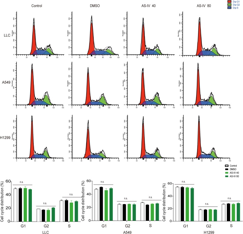
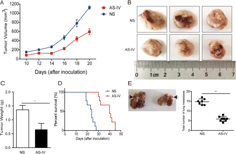
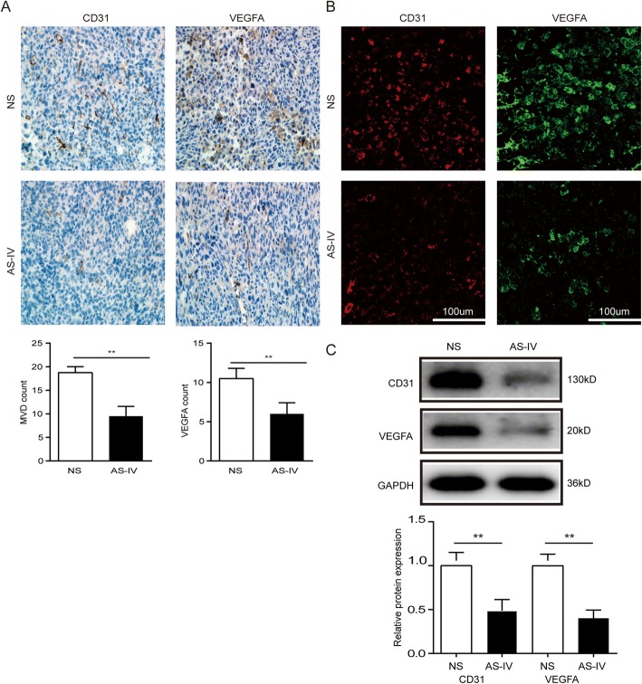
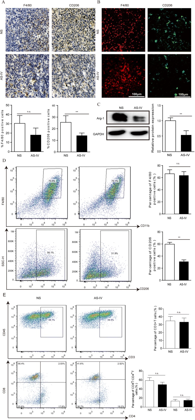
Results: Here we found that AS-IV significantly inhibited IL-13 and IL-4-induced M2 polarization of macrophages, as illustrated by reduced expression of CD206 and M2-associated genes, and that AS-IV suppressed the M2-CM-induced invasion, migration, and angiogenesis of A549 and H1299 cells. In vivo experiments demonstrated that AS-IV greatly inhibited tumor growth and reduced the number of metastases of Lewis lung cancer. The percentage of M2 macrophages was decreased in tumor tissue after AS-IV treatment. Furthermore, AS-IV inhibited AMPKα activation in M2 macrophages, and silencing of AMPKα partially abrogated the inhibitory effect of AS-IV.

Conclusions: AS-IV reduced the growth, invasion, migration, and angiogenesis of lung cancer by blocking the M2 polarization of macrophages partially through the AMPK signaling pathway, which appears to play an important role in AS-IV's ability to inhibit the metastasis of lung cancer.

Keywords: Astragaloside IV; Lung cancer; Macrophage polarization; Tumor-associated macrophages.


Conflict of interest statement

Ethics approval and consent to participate

The study was approved by the ethical review board of the Fudan University Animal Care and Use Committee (No.201500051).

Consent for publication

Not applicable.

Competing interests

The authors declare that they have no competing interests.

Publisher’s Note

Springer Nature remains neutral with regard to jurisdictional claims in published maps and institutional affiliations.

Figures

Fig. 1
Fig. 1
Human THP-1 monocyte differentiation into M1 and M2 macrophages. a The morphology of M0 (non-polarized THP-1 cells) and the expression of CD14 as measured by flow cytometry. b Confocal microscopy showing the distinct morphology of THP-1–derived M1 and M2 macrophages stained with DAPI (blue) and with antibodies against CD68 (green). c Flow cytometry demonstrated cell surface expression of CD86 and CD206 in the M0, M1, and M2 populations. d and e The mRNA expression levels of M1 and M2 macrophage markers were measured by QT-PCR. Data are presented as the mean ± SEM from three independent experiments. All results show the relative fold change compared to M0 (non-polarized THP-1 cells). *p < 0.05, **p < 0.01, and n.s. = no significance. IL, interleukin; TNF-α, tumor necrosis factor-α; iNOS, inducible nitric oxide synthase; CCR, chemokine receptor; CCL, C-C motif chemokine; MMP, matrix metalloproteinase
Fig. 2
Fig. 2
AS-IV inhibited macrophage M2 polarization. Macrophages derived from THP-1 cells were stimulated with IL-4/IL-13 with or without AS-IV (80 nM) for 48 h. a Flow cytometry was used to quantify the expression of CD206, an M2 macrophage marker, and CD86, an M1 marker. b QT-PCR was performed to detect gene levels in M2 and M1 macrophages. M2-associated genes included PPARγ, Arg-1, and CD206, and M1-associated genes included iNOS, TNF-α, and COX2. c Representative images of M0 and M2 macrophages with and without AS-IV stained with antibodies against Arg-1 (green) and with DAPI (blue). d The levels of IL-10 and TGF-β in the cell culture supernatants were measured by ELISA. Data are presented as the mean ± SEM from three independent experiments. Compared to M0, **p < 0.01, *p < 0.05, and n.s., no significance; compared to M2, ##p < 0.01, #p < 0.05, and n.s., no significance
Fig. 3
Fig. 3
AS-IV inhibited the migration and invasion of tumor cells. Macrophages derived from THP-1 were stimulated with IL-4/IL-13 with or without AS-IV (80 nM) for 48 h, and the conditioned medium (CM) was collected. A549 and H1299 cells were cultured with different CM. (a and b) The effect of AS-IV on A549 and H1299 invasion and migration was evaluated by wound scratch assay and trans-well assay, respectively. (c and d) The mRNA levels of genes associated with migration and angiogenesis in macrophages. Data are presented as the mean ± SEM from three independent experiments. Compared to M0, **p < 0.01, *p < 0.05, and n.s., no significance; compared to M2, ##p < 0.01, #p < 0.05, and n.s., no significance
Fig. 4
Fig. 4
AS-IV treatment inhibited AMPK signaling in the M2 polarization of macrophages. a Macrophages derived from THP-1 cells were stimulated with IL-4/IL-13 with or without AS-IV (80 nM) for 48 h. The levels of AMPKα and p-AMPKα as measured by western blot. b Macrophages derived from THP-1 cells were stimulated with IL-4/IL-13 with or without AS-IV (80 nM) for 12 h, 24 h, 36 h, and 48 h. AMPKα and p-AMPKα1 levels were measured by western blot. c The mRNA levels of AMPKα in macrophages were knocked down by siRNAs. d Macrophages were transiently transfected with siRNA 2 for 24 h, and AMPKα protein expression in transfected cells was analyzed by western blot. e siRNA2 blocked AMPKα expression. The percentage of CD206-positive macrophages was determined by flow cytometry. All experiments were repeated three times. Compared to M0, **p < 0.01, *p < 0.05, and n.s., no significance; compared to M2, ##p < 0.01, #p < 0.05, and n.s., no significance
Fig. 5
Fig. 5
AS-IV was not toxic to tumor cells. LLC, A549, and H1299 cells were treated with blank control, DMSO, 40 μM AS-IV, or 80 μM AS-IV for 48 h. The populations of cells in each phase of the cell cycle were detected by flow cytometry using PI staining. All of the data are presented as the mean ± SEM from three independent experiments
Fig. 6
Fig. 6
AS-IV inhibited the growth and migration of LLC tumors and prolonged survival in vivo. a The tumor volumes were determined at different time points. N = 6–15 animals per group. b Representative image of tumors at day 22. c The weights of isolated tumor tissues were measured. d Survival curves. e Representative image of lung metastases at day 22 and total number of lung metastases in the intravenous model. Data are presented as the mean ± SEM. N = 6–8 animals per group. Compared to the control group, **p < 0.01, *p < 0.05
Fig. 7
Fig. 7
AS-IV inhibited tumor vessel maturation. a–c The levels of CD31 and VEGFA were measured by IHC, immunofluorescence, and western blot. Data are presented as the mean ± SEM. N = 6–7 animals per group. Compared to the NS group, **p < 0.01
Fig. 8
Fig. 8
AS-IV suppressed M2 macrophage polarization in the subcutaneous mouse model. (a and b) The infiltrated macrophages in tumor tissues were stained with the macrophage marker F4/80 and the M2 marker CD206 by IHC and immunofluorescence, and the quantified data are shown. (c) Western blot analysis of Arg-1 expression in tumor tissues. (d) Macrophages from tumor tissue were stained with antibodies against F4/80 and CD206 and analyzed using flow cytometry. (e) T cells from tissues were stained with antibodies against CD3, CD4, and CD8 and assayed by flow cytometry. Data are presented as the mean ± SEM. N = 6–7 animals per group. Compared to the NS group, **p < 0.01, *p < 0.05, n.s., no significant difference

References

    1. Torre LA, et al. Global cancer statistics, 2012. CA Cancer J Clin. 2015;65(2):87–108. doi: 10.3322/caac.21262. - DOI - PubMed
    1. Conway EM, et al. Macrophages, inflammation, and lung Cancer. Am J Respir Crit Care Med. 2016;193(2):116–130. doi: 10.1164/rccm.201508-1545CI. - DOI - PubMed
    1. Doebele RC, et al. Mechanisms of resistance to crizotinib in patients with ALK gene rearranged non-small cell lung cancer. Clin Cancer Res. 2012;18(5):1472–1482. doi: 10.1158/1078-0432.CCR-11-2906. - DOI - PMC - PubMed
    1. Noone AM, et al. SEER Cancer Statistics Review. Bethesda, MD: National Cancer Institute; 1975-2015. https://seer.cancer.gov/csr/1975_2015/.
    1. Korniluk A, et al. From inflammation to cancer. Ir J Med Sci. 2017;186(1):57–62. doi: 10.1007/s11845-016-1464-0. - DOI - PMC - PubMed

MeSH terms