. 2018 Aug 29;37(1):202.
doi: 10.1186/s13046-018-0875-3.

SP1-induced lncRNA AGAP2-AS1 expression promotes chemoresistance of breast cancer by epigenetic regulation of MyD88

Affiliations

SP1-induced lncRNA AGAP2-AS1 expression promotes chemoresistance of breast cancer by epigenetic regulation of MyD88

Huaying Dong et al. J Exp Clin Cancer Res. .

Retraction in

Abstract

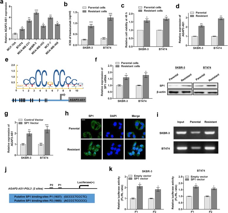
Background: Resistance to trastuzumab has become a leading cause of mortality in breast cancer patients and is one of the major obstacles for improving the clinical outcome. Cell behavior can be modulated by long non-coding RNAs (lncRNAs), but the contribution of lncRNAs in trastuzumab resistance to breast cancer is largely unknown. To this end, the involvement and regulatory function of lncRNA AGAP2-AS1 in human breast cancer are yet to be investigated.

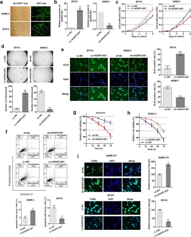
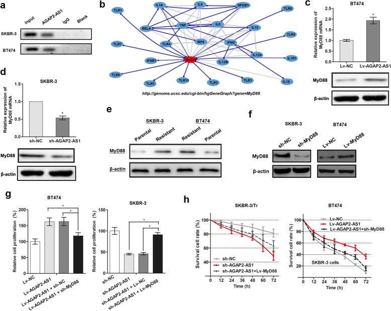
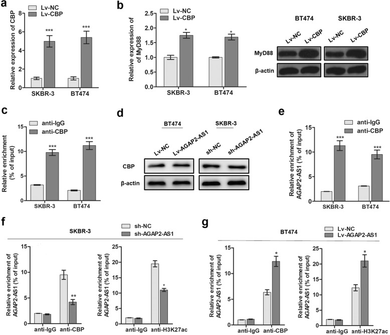
Methods: Trastuzumab-resistant SKBR-3 and BT474 cells were obtained by continuous culture with 5 mg/mL trastuzumab for 6 months. RT-qPCR assay was used to determine the expression of AGAP2-AS1 in tissues and cells. RNA fluorescence in situ hybridization was used to investigate the subcellular location of AGAP2-AS1 in breast cancer cells. Bioinformatic analysis, chromatin immunoprecipitation (ChIP), RNA immunoprecipitation (RIP), western blotting, and immunofluorescence were carried out to verify the regulatory interaction of AGAP2-AS1, CREB-binding protein (CBP), and MyD88. In addition, a series of in vitro assays and a xenograft tumor model were used to analyze the functions of AGAP2-AS1 in breast cancer cells.

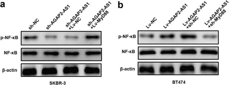
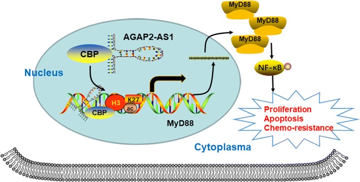
Results: AGAP2-AS1 was upregulated and transcriptionally induced by SP1 in breast cancer. Overexpression of AGAP2-AS1 promoted cell growth, suppressed apoptosis, and caused trastuzumab resistance, whereas knockdown of AGAP2-AS1 showed an opposite effect. MyD88 was identified as a downstream target of AGAP2-AS1 and mediated the AGAP2-AS1-induced oncogenic effects. Mechanistically, the RIP assay revealed that AGAP2-AS1 could bind to CBP, a transcriptional co-activator. ChIP assays showed that AGAP2-AS1-bound CBP increased the enrichment of H3K27ac at the promoter region of MyD88, thus resulting in the upregulation of MyD88. Gain- and loss-of-function assays confirmed that the NF-κB pathway was activated by MyD88 and AGAP2-AS1. Furthermore, high AGAP2-AS1 expression was associated with poor clinical response to trastuzumab therapy in breast cancer patients.

Conclusion: AGAP2-AS1 could promote breast cancer growth and trastuzumab resistance by activating the NF-κB signaling pathway and upregulating MyD88 expression. Therefore, AGAP2-AS1 may serve as a novel biomarker for prognosis and act as a therapeutic target for the trastuzumab treatment.

Keywords: Breast cancer; H3K27 acetylation; MyD88; SP1; Trastuzumab; lncRNA AGAP2-AS1.


Conflict of interest statement

Ethics approval and consent to participate

The present study was authorized by the Ethics Committee of Hainan General Hospital. All procedures performed in studies were in accordance with the ethical standards. All patients and volunteers were anonymous and have provided written informed consent.

Consent for publication

Not applicable.

Competing interests

The authors declare that they have no competing interests.

Publisher’s Note

Springer Nature remains neutral with regard to jurisdictional claims in published maps and institutional affiliations.

Figures

Fig. 1
Fig. 1
LncRNA AGAP2-AS1 is upregulated and induced by SP1 in breast cancer cells. a RT-qPCR was performed to detect the expression level of AGAP2-AS1 in breast cancer cells. b The IC50 value of trastuzumab was detected for both sensitive and resistant cells by cell viability assay. c The cell viability of both trastuzumab-resistant and sensitive cells were determined. d AGAP2-AS1 expression was identified by RT-qPCR in both trastuzumab-resistant and sensitive cells. e Prediction of SP1 binding site in the AGAP2-AS1 promoter region using JASPAR (http://jaspar.genereg.net/). f The expression of SP1 in gefitinib resistant and parental cells at transcript (left panel) and protein (right panel) levels. g RT-qPCR analysis of AGAP2-AS1 expression after SP1 was overexpressed. h FISH analysis of the enriched level of SP1 gene in nucleus of the SKBR-3 or SKBR-3/Tr cells. i ChIP assay was performed to detect the relative enrichment of SP1 in promoter of AGAP2-AS1. j Schematic presentation of SP1 binding sites in the promoter region of AGAP2-AS1. k Luciferase activity analysis of the two binding sites in cells transfected with respective oligonucleotides. *p < 0.05, **p < 0.01, ***p < 0.001
Fig. 2
Fig. 2
AGAP2-AS1 promotes proliferation and suppresses apoptosis of breast cancer cells. a The oligonucleotides labeled with GFP green fluorescence were transfected as described in the methods. b Transfection efficiency was identified by detecting AGAP2-AS1 expression via RT-qPCR. c MTT assay showed the functional effect of AGAP2-AS1 on cell proliferation of breast cancer cells. d Colony formation assay was used to determine the functional role of AGAP2-AS1. e Immunofluorescence analysis of Ki-67 expression in breast cancer cells after infection with respective oligonucleotides. f FACS apoptosis analysis of cells with overexpression or knockdown of AGAP2-AS1. g, h Cell viability was evaluated in cells treated with trastuzumab (0.5 mg/mL) after infection of respective oligonucleotides. i TUNEL assay was used to determine the function of AGAP2-AS1 for trastuzumab-induced cell apoptosis. *p < 0.05, **p < 0.01, ***p < 0.001
Fig. 3
Fig. 3
MyD88 is a downstream target of AGAP2-AS1 function in breast cancer. a RNA pull-down assay was performed to verify the enrichment of MyD88 by AGAP2-AS1 in the breast cancer cells. b Schematic diagram of the network of MyD88 based on ENCODE database (http://genome.ucsc.edu). c, d MyD88 was positively regulated by AGAP2-AS1 at both transcript and protein levels in SKBR-3 and BT474 cells. e MyD88 expression was determined in the trastuzumab-resistant and the parental breast cancer cells by western blot experiment. f MyD88 expression was detected via western blot after transfection of sh-MyD88 or Lv-MyD88. g Knockdown of MyD88 dramatically abrogated the effects of Lv-AGAP2-AS1 on cell proliferation whereas overexpression of MyD88 reversed the effect induced by sh-AGAP2-AS1. h Co-transfection of Lv-MyD88 reversed the effects of AGAP2-AS1 knockdown in SKBR-3/Tr cells whereas co-expression of sh-MyD88 abrogated the resistance induced by Lv-AGAP2-AS1 in the BT474 parental cells. *p < 0.05
Fig. 4
Fig. 4
AGAP2-AS1 promotes the expression of MyD88 by modifying H3K27 acetylation. a Bioinformatics analysis showed that the promoter of MyD88 had a high enrichment of H3K27ac. b ChIP assay was used to verify the enrichment of H3K27ac in MyD88 promoter in the breast cancer cells and the normal breast epithelial cells. c ChIP assay showed that the enrichment of H3K27ac was higher in the trastuzumab resistant cells than in the parental cells. d MyD88 expression was detected by western blot in breast cancer cells treated with C646 or DMSO. e FISH analysis of the subcellular location of AGAP2-AS1 with a specific probe in breast cancer cells. f ChIP assay showed that AGAP2-AS1 positively regulated the enrichment of H3K27ac at the MyD88 promoter. *p < 0.05, ***p < 0.001
Fig. 5
Fig. 5
AGAP2-AS1 mediates H3K27 acetylation at the promoter of MyD88 by binding with CBP. a CBP expression was detected by RT-qPCR. b The influence of CBP on MyD88 expression was identified by RT-qPCR (left panel) and western blot (right panel). c ChIP assay by using CBP antibody tested the enrichment at MyD88 promoter. d Western blot analysis indicated that AGAP2-AS1 showed little effect on CBP expression. e RIP experiment showed that anti-CBP antibody could precipitate AGAP2-AS1 in SKBR-3 and BT474 cells. f, g ChIP-qPCR revealed that AGAP2-AS1 influenced the enrichment of CBP and the level of H3K27ac acetylation at the promoter region of MyD88. *p < 0.05, **p < 0.01, ***p < 0.001
Fig. 6
Fig. 6
AGAP2-AS1 facilitates trastuzumab resistance via MyD88 in vivo. a Images of tumors that developed in xenograft transplanted nude mouse tumor models treated by administering 5 mg/kg trastuzumab or PBS (for control group) intraperitoneally once every 2 days for 3 weeks in different groups. Four treatment groups of mice xenograft were established: Group I (Lv-NC-transfected cells + PBS), Group II (Lv-AGAP2-AS1-transfected cells + PBS), Group III (Lv-NC-transfected cells + trastuzumab treatment) and Group IV (Lv- AGAP2-AS1-transfected cells + trastuzumab treatment). Six mice were included in each group and more than three mice remained after the treatment regimen excluding mice that were dead or with complications, such as skin necrosis due to infection. b Weights of tumors that developed in xenografts from different groups are shown. c IHC analysis of expression levels of MyD88 in respective groups. d Images of tumors that developed in xenograft transplanted nude mouse tumor models treated orally with 5 mg/kg gefitinib once daily for 3 weeks in different transfection groups. *p < 0.05, **p < 0.01
Fig. 7
Fig. 7
NF-κB signaling pathways is promoted by the upregulation of MyD88 induced by AGAP2-AS1. a Western blot was performed to detect the activity of NF-κB in AGAP2-AS1-silenced SKBR-3 cells transfected with Lv-MyD88. b Western blot was performed to detect the activity of NF-κB in AGAP2-AS1-overexpressing BT474 cells transfected with sh-MyD88
Fig. 8
Fig. 8
AGAP2-AS1 expression is associated with chemoresponse to trastuzumab therapy in patients with breast cancer. a The expressions of AGAP2-AS1 and MyD88 in 42 paired breast cancer tissues. b The statistically significant association between AGAP2-AS1 and MyD88 expression in breast cancer specimens. c AGAP2-AS1 expression was determined via RT-qPCR for the trastuzumab-resistant and sensitive patients. d ROC curve was established to show the ability of AGAP2-AS1 in differentiating responding patients from non-responding patients. e The proportion of patients that showed resistance to trastuzumab therapy was significantly higher in high AGAP2-AS1 expressing groups than in low expression group. ***p < 0.001
Fig. 9
Fig. 9
A schematic diagram representing the role of AGAP2-AS1 in breast cancer chemoresistance via upregulating MyD88 expression through modifying H3K27 acetylation

References

    1. Torre LA, Bray F, Siegel RL, Ferlay J, Lortet-Tieulent J, Jemal A. Global cancer statistics, 2012. CA Cancer J Clin. 2015;65(2):87–108. doi: 10.3322/caac.21262. - DOI - PubMed
    1. Gonzalez-Angulo AM, Morales-Vasquez F, Hortobagyi GN. Overview of resistance to systemic therapy in patients with breast cancer. Adv Exp Med Biol. 2007;608:1–22. doi: 10.1007/978-0-387-74039-3_1. - DOI - PubMed
    1. Vu T, Claret FX. Trastuzumab: updated mechanisms of action and resistance in breast cancer. Front Oncol. 2012;2:62. doi: 10.3389/fonc.2012.00062. - DOI - PMC - PubMed
    1. Robidoux A, Tang G, Rastogi P, Geyer CE, Jr, Azar CA, Atkins JN, Fehrenbacher L, Bear HD, Baez-Diaz L, Sarwar S, et al. Lapatinib as a component of neoadjuvant therapy for HER2-positive operable breast cancer (NSABP protocol B-41): an open-label, randomised phase 3 trial. Lancet Oncol. 2013;14(12):1183–1192. doi: 10.1016/S1470-2045(13)70411-X. - DOI - PubMed
    1. Narayan M, Wilken JA, Harris LN, Baron AT, Kimbler KD, Maihle NJ. Trastuzumab-induced HER reprogramming in “resistant” breast carcinoma cells. Cancer Res. 2009;69(6):2191–2194. doi: 10.1158/0008-5472.CAN-08-1056. - DOI - PubMed

Publication types

MeSH terms