. 2018 Aug 29;49(1):83.
doi: 10.1186/s13567-018-0575-1.

A G1-lineage H9N2 virus with oviduct tropism causes chronic pathological changes in the infundibulum and a long-lasting drop in egg production

Affiliations

A G1-lineage H9N2 virus with oviduct tropism causes chronic pathological changes in the infundibulum and a long-lasting drop in egg production

Francesco Bonfante et al. Vet Res. .

Abstract

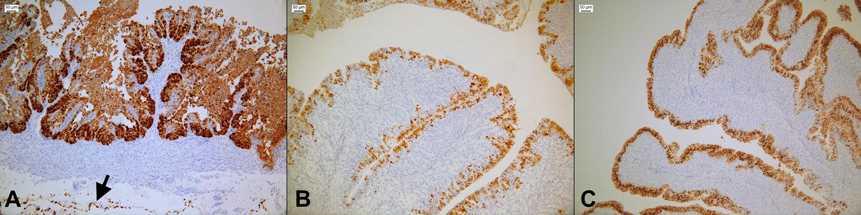
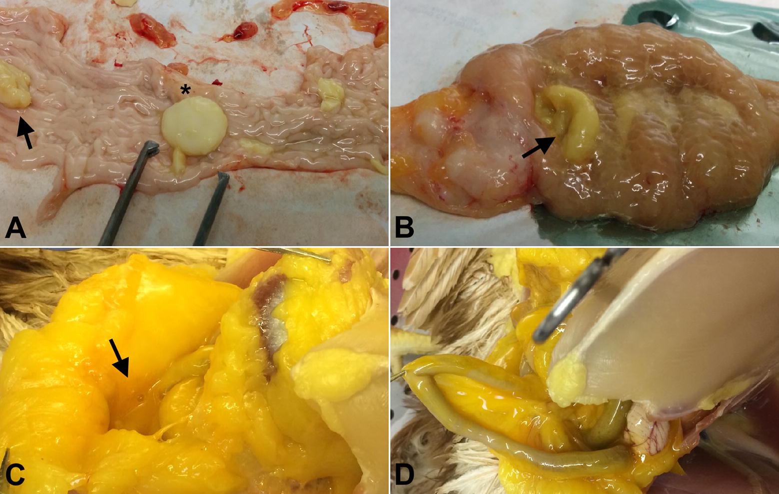
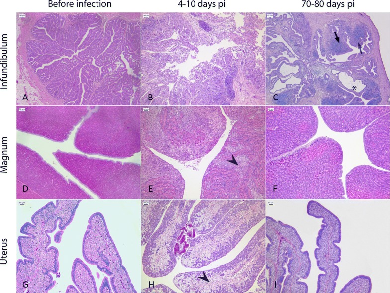
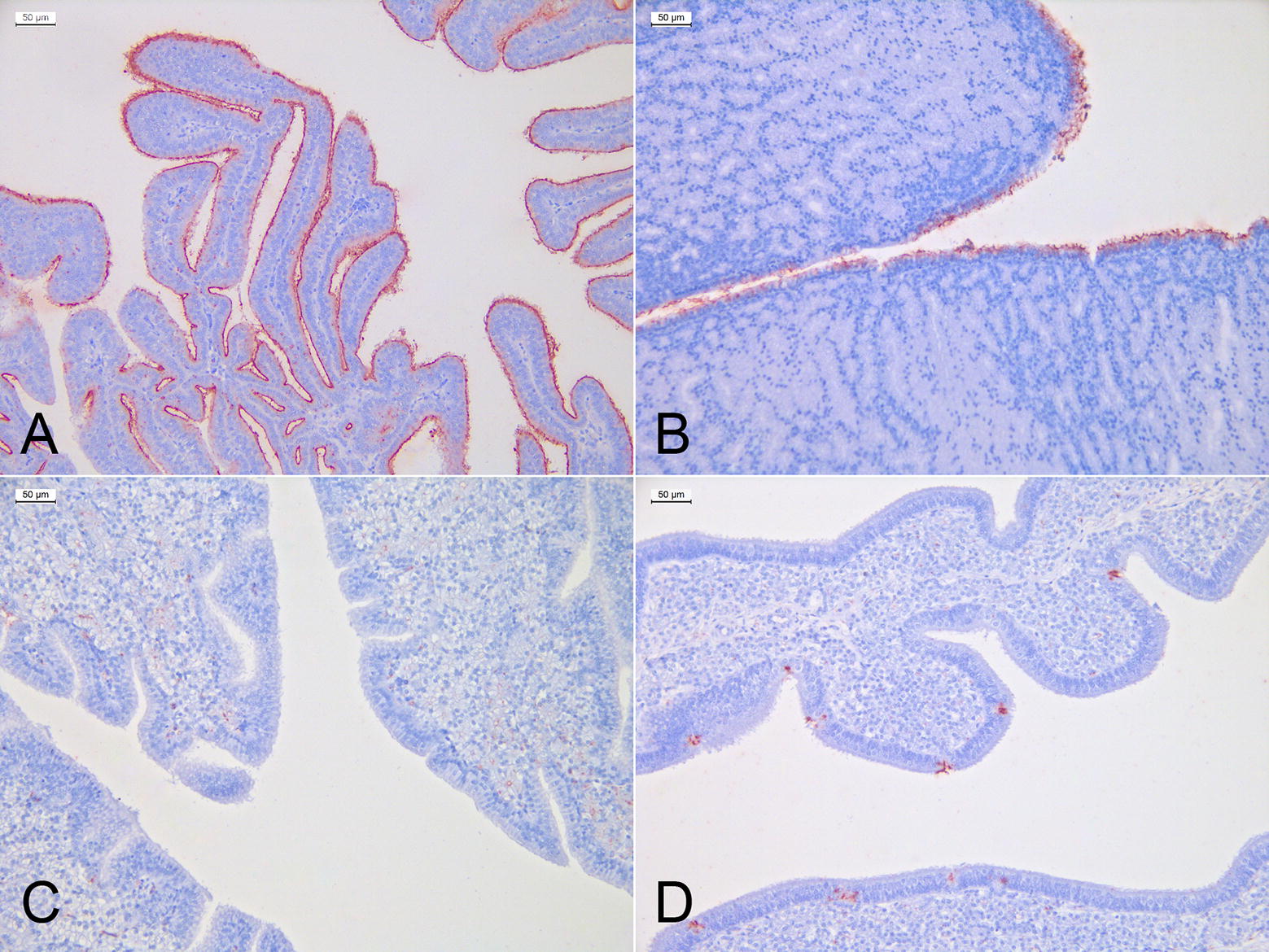
Since 1997, G1-lineage H9N2 avian influenza viruses have been circulating in Asia and later on in the Middle East, and they have been associated to mild respiratory disease, drops in egg production and moderate mortality in chickens, in particular in the presence of concurrent infections. In this study, we investigated the importance of the G1-lineage H9N2 A/chicken/Israel/1163/2011 virus as a primary pathogen in layers, analyzing its tropism and binding affinity for the oviduct tissues, and investigating the long-term impact on egg production. Besides causing a mild respiratory infection, the virus replicated in the oviduct of 60% of the hens causing different degrees of salpingitis throughout the organ, in particular at the level of the infundibulum, where the detection of the virus was associated with severe heterophilic infiltrate, and necrosis of the epithelium. Binding affinity assays confirmed that the infundibulum was the most receptive region of the oviduct. The drop in egg production was at its peek at 2 weeks post-infection (pi) (60% decrease) and continued up to 80 days pi (35% decrease). On day 80 pi, non-laying birds showed egg yolk peritonitis, and histopathological analyses described profound alteration of the infundibulum architecture, duct ectasia and thinning of the epithelium, while the rest of the oviduct and ovary appeared normal. Our results show that this H9N2 virus is a primary pathogen in layer hens, and that its replication in the infundibulum is responsible for acute and chronic lesions that limits the effective functionality of the oviduct, compromising the commercial life of birds.


Figures

Figure 1
Figure 1
Laying performance of challenged and control birds in experiment 1. Eggs were collected throughout the experiment, three times a day, keeping handling of animals to the minimum, in order to avoid the occurrence of stressful events. We compared the egg production between challenged and control birds on a weekly basis, adopting the Wilcoxon matched-pairs signed-ranks on daily mean values for each group. Significance level was set at P < 0.05. Data are shown here as mean ± standard deviations. The differences in the weekly egg production between the two groups were significant (P < 0.05) for 9 weeks after the challenge.
Figure 2
Figure 2
Virus loads in organs collected from birds in experiment 2, on days 4, 6 and 10 pi. Virus colonization of organs was assessed and quantified by means of qRRT-PCR and TCID50, in MDCK cells. Colored and black columns represent the number of virus RNA copies and TCID50/0.1 g of tissue, respectively. Limits of detections for each scale are indicated by lateral bars. Values are shown on an individual basis, as indicated by the bird ID number on the X axis. LOD: limit of detection, inf: infundibulum, ma: magnum, is: isthmus, ut: uterus.
Figure 3
Figure 3
Immunohistochemistry of the infundibulum, magnum and uterus of challenged birds in experiment 2. Representative 3-µm sections of oviducts collected during the acute phase of infection, in experiment 1. Sections were incubated with a monoclonal antibody targeting the Influenza A virus nucleoprotein. Positivity was revealed with 3, 3-diaminobenzidine (DAB), forming a brown precipitate. A Strong positivity of an infundibulum collected on day 4 pi, showing severe necrosis and erosion of the epithelial mucosa. The arrow indicates positivity of the mesenchymal cells lining the serosa. Epithelial staining of B magnum and C uterus collected on days 6 pi.
Figure 4
Figure 4
Gross pathology of the oviduct and abdomen. A A magnum collected on day 4 pi A defective egg and a fibrinous clot are indicated by an asterisk and an arrow, respectively. B A uterus collected on day 6 pi. The arrow indicates mucus and fibrin. C, D Egg yolk material in the abdomen of a bird necropsied on days 18 (C) and 80 pi (D).
Figure 5
Figure 5
Representative histological pictures of the infundibulum, magnum and uterus before, during and after the infection. AI All pictures were taken using a 10× objective from 4-μm sections stained with hematoxylin and eosin. B During the acute phase of infection (4–10 days pi), replication of the virus in the infundibulum was associated with the occurrence of severe necrosis, edema and granulomatous processes in the mucosa of the organ. C In the chronic phase of the disease (70–80 days pi), birds with egg yolk peritonitis showed a profound alteration of the infundibulum architecture due to the presence of ectatic glands (asterisk), hyperplastic lymphocytic aggregates (arrows) and thinning of the mucosa. E, H Lesions in the magnum and uterus were rarely observed during the acute phase of the infection and were limited to heterophilic necrosis and edema (arrow heads), respectively. F, I A full restoration of the tissue integrity was observed for the magnum and uterus in all subjects, after 70–80 days from infection.
Figure 6
Figure 6
Virus-histochemistry of the oviduct. Red staining indicates the binding of the FITC-labelled virus to the tissues. Sections are counterstained with hematoxylin. A The infundibulum showed a highly specific and uniform positivity of the cilia at the level of the epithelium lining the fimbriae. B In the magnum, we observed a specific staining of the ciliated epithelium but intensity was lower compared with the infundibulum. C No attachment of the virus was observed at the level of the isthmus epithelium. D The uterus showed a scattered but specific attachment of the virus to the cytoplasm of epithelial goblet cells.

References

    1. Swayne DE. Animal influenza. Hoboken: Wiley; 2016.
    1. Fusaro A, Monne I, Salviato A, Valastro V, Schivo A, Amarin NM, Gonzalez C, Ismail MM, Al-Ankari A-R, Al-Blowi MH, Khan OA, Maken Ali AS, Hedayati A, Garcia Garcia J, Ziay GM, Shoushtari A, Al Qahtani KN, Capua I, Holmes EC, Cattoli G. Phylogeography and evolutionary history of reassortant H9N2 viruses with potential human health implications. J Virol. 2011;85:8413–8421. doi: 10.1128/JVI.00219-11. - DOI - PMC - PubMed
    1. Davidson I, Fusaro A, Heidari A, Monne I, Cattoli G. Molecular evolution of H9N2 avian influenza viruses in Israel. Virus Genes. 2014;48:457–463. doi: 10.1007/s11262-014-1037-0. - DOI - PubMed
    1. Houadfi MEL, Fellahi S, Nassik S, Guérin J-L, Ducatez MF. First outbreaks and phylogenetic analyses of avian influenza H9N2 viruses isolated from poultry flocks in Morocco. Virol J. 2016;13:140. doi: 10.1186/s12985-016-0596-1. - DOI - PMC - PubMed
    1. Aouini R, Laamiri N, Ghram A. Novel gene mutations in Tunisian isolate of avian H9N2 influenza virus. J Vet Sci Technol. 2016;8:1–9. doi: 10.4172/2157-7579.1000405. - DOI