. 2018 Sep 20;131(18):jcs220251.
doi: 10.1242/jcs.220251.

The Drosophila Epidermal Growth Factor Receptor does not act in the nucleus

Affiliations

The Drosophila Epidermal Growth Factor Receptor does not act in the nucleus

Maximilien Courgeon et al. J Cell Sci. .

Abstract

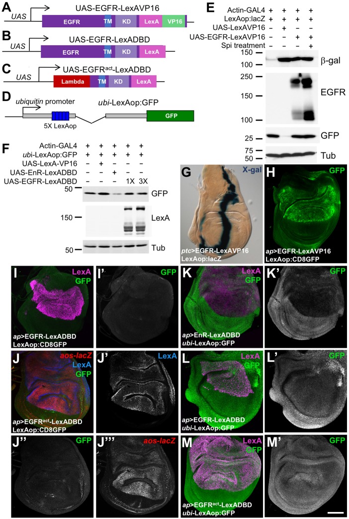
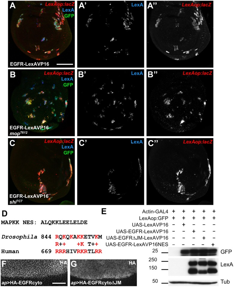
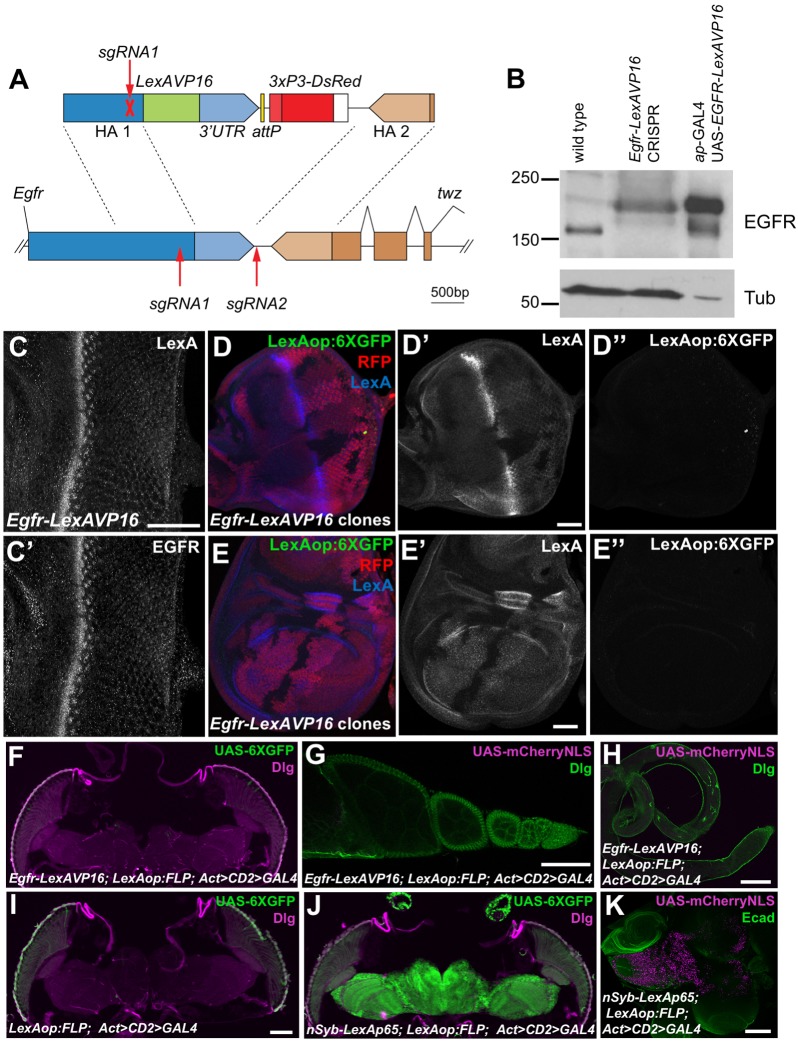
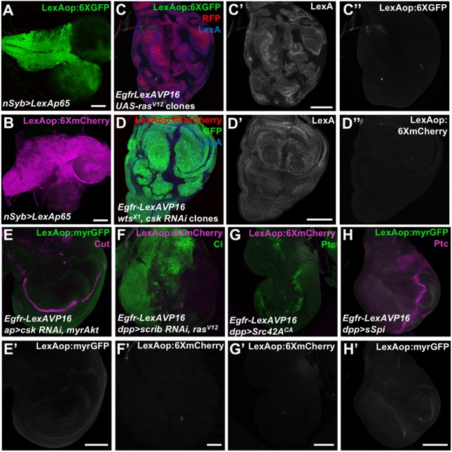
Mammalian members of the ErbB family, including the epidermal growth factor receptor (EGFR), can regulate transcription, DNA replication and repair through nuclear entry of either the full-length proteins or their cleaved cytoplasmic domains. In cancer cells, these nuclear functions contribute to tumor progression and drug resistance. Here, we examined whether the single Drosophila EGFR can also localize to the nucleus. A chimeric EGFR protein fused at its cytoplasmic C-terminus to DNA-binding and transcriptional activation domains strongly activated transcriptional reporters when overexpressed in cultured cells or in vivo However, this activity was independent of cleavage and endocytosis. Without an exogenous activation domain, EGFR fused to a DNA-binding domain did not activate or repress transcription. Addition of the same DNA-binding and transcriptional activation domains to the endogenous Egfr locus through genome editing led to no detectable reporter expression in wild-type or oncogenic contexts. These results show that, when expressed at physiological levels, the cytoplasmic domain of the Drosophila EGFR does not have access to the nucleus. Therefore, nuclear EGFR functions are likely to have evolved after vertebrates and invertebrates diverged.

Keywords: Cancer; EGFR; Nucleus; Transcription.


Conflict of interest statement

Competing interestsThe authors declare no competing or financial interests.

Figures

Fig. 1.
Fig. 1.
The EGFR activates transcription when fused to DNA-binding and activation domains. (A–D) Diagrams of UAS-EGFRLexAVP16 (A), UAS-EGFRLexADBD (B), UAS-EGFRactLexADBD (C) and ubi-LexAop:GFP (D). TM, transmembrane domain; KD, kinase domain; Lambda, dimerization domain. (E,F) Western blots of lysates from S2R+ cells transfected as indicated, and either treated for 4.5 h with purified sSpiCS or untreated. In E, the β-galactosidase (β-gal) blot reflects expression from LexAop:lacZ. The EGFR blot shows full-length EGFR-LexAVP16 (upper bands) and its cleaved cytoplasmic domain (lower bands). Co-transfected UAS-GFP was used to normalize for transfection efficiency and β-Tubulin (Tub) for total protein. Toxicity of UAS-LexAVP16 resulted in fewer surviving GFP-expressing cells, each of which strongly expressed LexAop:lacZ. In F, the GFP blot reflects ubi-LexAop:GFP reporter expression, which is reduced by transfection of UAS-EnR-LexADBD but not by equal or 3-fold higher amounts of UAS-EGFR-LexADBD. (G,H) Wing discs expressing UAS-EGFRLexAVP16 at the anterior-posterior compartment boundary with patched (ptc)-GAL4 (G) or in the dorsal compartment with ap-GAL4 (H), stained with X-gal, reflecting LexAop:lacZ expression (G) or anti-GFP antibody reflecting LexAop:CD8GFP (H). EGFR–LexAVP16 strongly activates both reporters. (I–M) Wing discs from flies in which ap-GAL4 drove UAS-EGFRLexADBD (I,L), UAS-EGFRactLexADBD (J,M) or UAS-EnRLexADBD (K). Anti-LexA antibody staining (J′, blue in J, magenta in I,K–M) shows the expression of these constructs. Anti-GFP antibody staining (I′,J″,K′–M′, green in K–M) reflects expression from LexAop:GFP (I,J) or ubi-LexAop:GFP (K–M). Anti-β-gal antibody staining shows aos-lacZ expression (J‴, red in J). Scale bar (M′): 100 μm. Fusion of the LexA DNA-binding domain to wild-type or activated EGFR (EGFRact) does not enable it to activate or repress these reporters, although EnRLexADBD represses ubi-LexAop:GFP.
Fig. 2.
Fig. 2.
Overexpressed EGFR–LexAVP16 can bypass normal trafficking. (A–C) Wing discs in which UAS-EGFR-LexAVP16 is expressed in clones of cells that are wild type (A), mop mutant (B) or shi mutant (C). Clones are marked by anti-GFP antibody staining (green) and anti-LexA antibody staining (A′–C′, blue). Anti-β-gal antibody staining reflects LexAop:lacZ expression (A″–C″, red), which is not reduced by the blocking of EGFR cleavage that occurs in mop cells or of endocytosis that occurs in shi cells. (D) Sequence of the NES from MAPKK and an alignment of the amino acids immediately following the transmembrane domains of Drosophila and human EGFR. Basic residues are in red. (E) Western blots of S2R+ cell lysates transfected with the indicated constructs. Deleting the juxtamembrane region reduces EGFR–LexAVP16 cleavage, but neither this deletion nor adding an NES reduces reporter expression. (F,G) Portions of wing discs spanning the dorsal-ventral boundary in which UAS-HA-EGFRcyto (F) or UAS-HA-EGFRcytoΔJM (G) is expressed in the dorsal (upper) compartment with ap-GAL4. Anti-HA antibody staining shows cytoplasmic localization. Scale bars: 50 μm (A), 20 μm (F).
Fig. 3.
Fig. 3.
EGFR–LexAVP16 does not activate transcription when expressed at physiological levels. (A) The sgRNAs used to edit the endogenous Egfr sequence (lower) and the construct used for homology-directed repair (upper). (B) Western blot of lysates from wild-type and homozygous Egfr-LexAVP16 CRISPR adult flies and from ap-GAL4; UAS-EGFRLexAVP16 larval wing discs. In the CRISPR flies, EGFR is expressed at wild-type levels and migrates at the size for EGFR–LexAVP16. The wing disc extracts had much stronger EGFR–LexAVP16 expression and less protein was loaded. (C) Egfr-LexAVP16 eye disc stained with anti-LexA (C) and anti-EGFR (C′) antibody. Scale bar: 50 μm. (D,E) Eye disc (D) and wing disc (E) with clones lacking the Egfr-LexAVP16-modified locus marked by the absence of RFP (red) and LexA (D′,E′, blue). Anti-GFP antibody staining shows LexAop:6XGFP expression (D″,E″, green), which is not detectable in cells with Egfr-LexAVP16 above the background staining present in wild-type cells. Scale bars: 50 μm. (F–H) Egfr-LexAVP16; LexAop:FLP; Act>CD2>GAL4 adult head (F), ovary (G) and testis (H) showing no expression of UAS-6×GFP stained with anti-GFP antibody (green, F) or of UAS-mCherry-NLS (magenta, G,H). (I) A negative control adult head without Egfr-LexAVP16. (J,K) nSyb-LexAp65; LexAop:FLP; Act>CD2>GAL4 induces strong expression of UAS-6×GFP in adult brain (J) and of UAS-mCherry-NLS in larval brain (K). F–J are stained for Discs large (Dlg, magenta in F,I,J; green in G,H) and K is stained for E-cadherin (Ecad, green). Scale bars: 100 μm.
Fig. 4.
Fig. 4.
Oncogenic transformation does not induce EGFR nuclear localization. (A,B) Larval brains showing LexAop:6XGFP (anti-GFP antibody, A) and LexAop:6XmCherry (B) expression driven by nSyb>LexAp65. (C) Wing disc with UAS-rasV12 expressed in clones homozygous for Egfr-LexAVP16, labeled with RFP (red), showing LexA (C′, blue) and LexAop:6XGFP (C″, green). (D) Egfr-LexAVP16 wing disc with wts mutant clones expressing UAS-csk RNAi and GFP (green), showing LexA (D′, blue) and LexAop:6XmCherry (D″, red) expression. (E–H) Egfr-LexAVP16 wing discs expressing UAS-csk RNAi and UAS-myrAkt (E), UAS-scrib RNAi and UAS-rasV12 (F), UAS-Src42ACA (G) or UAS-sSpi (H) in the dorsal compartment with ap-GAL4 (E) or at the anterior-posterior compartment boundary with decapentaplegic (dpp)-GAL4 (F-H), showing LexAop:myrGFP (anti-GFP, E′,H′, green in E,H) or LexAop:6XmCherry (F′,G′, magenta in F,G) expression. Cut (magenta in E) marks the dorsal-ventral boundary, Cubitus interruptus (Ci, green in F) marks the anterior compartment, and Ptc (green in G, magenta in H) marks the anterior-posterior boundary. None of these oncogenic conditions induce reporter activation by Egfr-LexAVP16. Scale bars: 100 μm.

References

    1. Adilakshmi T., Ness-Myers J., Madrid-Aliste C., Fiser A. and Tapinos N. (2011). A nuclear variant of ErbB3 receptor tyrosine kinase regulates ezrin distribution and Schwann cell myelination. J. Neurosci. 31, 5106-5119. 10.1523/JNEUROSCI.5635-10.2011 - DOI - PMC - PubMed
    1. Beguelin W., Diaz Flaque M. C., Proietti C. J., Cayrol F., Rivas M. A., Tkach M., Rosemblit C., Tocci J. M., Charreau E. H., Schillaci R. et al. (2010). Progesterone receptor induces ErbB-2 nuclear translocation to promote breast cancer growth via a novel transcriptional effect: ErbB-2 function as a coactivator of Stat3. Mol. Cell. Biol. 30, 5456-5472. 10.1128/MCB.00012-10 - DOI - PMC - PubMed
    1. Bitler B. G., Goverdhan A. and Schroeder J. A. (2010). MUC1 regulates nuclear localization and function of the epidermal growth factor receptor. J. Cell Sci. 123, 1716-1723. 10.1242/jcs.062661 - DOI - PMC - PubMed
    1. Brand T. M., Iida M., Luthar N., Starr M. M., Huppert E. J. and Wheeler D. L. (2013). Nuclear EGFR as a molecular target in cancer. Radiother. Oncol. 108, 370-377. 10.1016/j.radonc.2013.06.010 - DOI - PMC - PubMed
    1. Carpenter G. and Liao H.-J. (2013). Receptor tyrosine kinases in the nucleus. Cold Spring Harb. Perspect. Biol. 5, a008979 10.1101/cshperspect.a008979 - DOI - PMC - PubMed

Publication types

LinkOut - more resources