

Comparative In Vitro and In Vivo Analysis of H1N1 and H1N2 Variant Influenza Viruses Isolated from Humans between 2011 and 2016
- PMID: 30158292
- PMCID: PMC6206486
- DOI: 10.1128/JVI.01444-18
Comparative In Vitro and In Vivo Analysis of H1N1 and H1N2 Variant Influenza Viruses Isolated from Humans between 2011 and 2016
Abstract
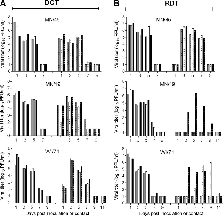
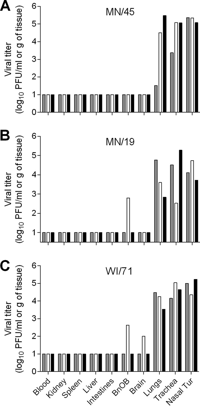
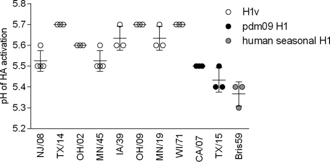
Influenza A virus pandemics are rare events caused by novel viruses which have the ability to spread in susceptible human populations. With respect to H1 subtype viruses, swine H1N1 and H1N2 viruses occasionally cross the species barrier to cause human infection. Recently isolated from humans (termed variants), swine viruses were shown to display great genetic and antigenic diversity, hence posing considerable public health risk. Here, we utilized in vitro and in vivo approaches to provide characterization of H1 subtype variant viruses isolated since the 2009 pandemic and discuss the findings in context with previously studied H1 subtype human isolates. The variant viruses were well adapted to replicate in the human respiratory cell line Calu-3 and the respiratory tracts of mice and ferrets. However, with respect to hemagglutinin (HA) activation pH, the variant viruses had fusion pH thresholds closer to that of most classical swine and triple-reassortant H1 isolates rather than viruses that had adapted to humans. Consistent with previous observations for swine isolates, the tested variant viruses were capable of efficient transmission between cohoused ferrets but could transmit via respiratory droplets to differing degrees. Overall, this investigation demonstrates that swine H1 viruses that infected humans possess adaptations required for robust replication and, in some cases, efficient respiratory droplet transmission in a mammalian model and therefore need to be closely monitored for additional molecular changes that could facilitate transmission among humans. This work highlights the need for risk assessments of emerging H1 viruses as they continue to evolve and cause human infections.IMPORTANCE Influenza A virus is a continuously evolving respiratory pathogen. Endemic in swine, H1 and H3 subtype viruses sporadically cause human infections. As each zoonotic infection represents an opportunity for human adaptation, the emergence of a transmissible influenza virus to which there is little or no preexisting immunity is an ongoing threat to public health. Recently isolated variant H1 subtype viruses were shown to display extensive genetic diversity and in many instances were antigenically distinct from seasonal vaccine strains. In this study, we provide characterization of representative H1N1v and H1N2v viruses isolated since the 2009 pandemic. Our results show that although recent variant H1 viruses possess some adaptation markers of concern, these viruses have not fully adapted to humans and require further adaptation to present a pandemic threat. This investigation highlights the need for close monitoring of emerging variant influenza viruses for molecular changes that could facilitate efficient transmission among humans.
Keywords: H1N1; H1N2; ferret; influenza; pathogenesis; risk assessment; variant virus.
Copyright © 2018 American Society for Microbiology.
Figures


Similar articles
-
Antigenically Diverse Swine Origin H1N1 Variant Influenza Viruses Exhibit Differential Ferret Pathogenesis and Transmission Phenotypes.J Virol. 2018 May 14;92(11):e00095-18. doi: 10.1128/JVI.00095-18. Print 2018 Jun 1. J Virol. 2018. PMID: 29540597 Free PMC article.
-
Pathogenesis and Transmission of Genetically Diverse Swine-Origin H3N2 Variant Influenza A Viruses from Multiple Lineages Isolated in the United States, 2011-2016.J Virol. 2018 Jul 31;92(16):e00665-18. doi: 10.1128/JVI.00665-18. Print 2018 Aug 15. J Virol. 2018. PMID: 29848587 Free PMC article.
-
Evaluation of the zoonotic potential of a novel reassortant H1N2 swine influenza virus with gene constellation derived from multiple viral sources.Infect Genet Evol. 2015 Aug;34:378-93. doi: 10.1016/j.meegid.2015.06.005. Epub 2015 Jun 4. Infect Genet Evol. 2015. PMID: 26051886
-
[Swine influenza virus: evolution mechanism and epidemic characterization--a review].Wei Sheng Wu Xue Bao. 2009 Sep;49(9):1138-45. Wei Sheng Wu Xue Bao. 2009. PMID: 20030049 Review. Chinese.
-
Swine influenza viruses: an Asian perspective.Curr Top Microbiol Immunol. 2013;370:147-72. doi: 10.1007/82_2011_195. Curr Top Microbiol Immunol. 2013. PMID: 22266639 Review.
Cited by
-
Pathogenesis and transmission of human seasonal and swine-origin A(H1) influenza viruses in the ferret model.Emerg Microbes Infect. 2022 Dec;11(1):1452-1459. doi: 10.1080/22221751.2022.2076615. Emerg Microbes Infect. 2022. PMID: 35537045 Free PMC article.
-
Identification, Genetic Analysis, and Pathogenicity of Classical Swine H1N1 and Human-Swine Reassortant H1N1 Influenza Viruses from Pigs in China.Viruses. 2020 Jan 2;12(1):55. doi: 10.3390/v12010055. Viruses. 2020. PMID: 31906591 Free PMC article.
-
Machine Learning Methods for Predicting Human-Adaptive Influenza A Viruses Based on Viral Nucleotide Compositions.Mol Biol Evol. 2020 Apr 1;37(4):1224-1236. doi: 10.1093/molbev/msz276. Mol Biol Evol. 2020. PMID: 31750915 Free PMC article.
-
Kinetics and magnitude of viral RNA shedding as indicators for Influenza A virus transmissibility in ferrets.Commun Biol. 2023 Jan 23;6(1):90. doi: 10.1038/s42003-023-04459-0. Commun Biol. 2023. PMID: 36690690 Free PMC article.
-
Aerosol Transmission from Infected Swine to Ferrets of an H3N2 Virus Collected from an Agricultural Fair and Associated with Human Variant Infections.J Virol. 2020 Jul 30;94(16):e01009-20. doi: 10.1128/JVI.01009-20. Print 2020 Jul 30. J Virol. 2020. PMID: 32522849 Free PMC article.
References
-
- Simonsen L, Spreeuwenberg P, Lustig R, Taylor RJ, Fleming DM, Kroneman M, Van Kerkhove MD, Mounts AW, Paget WJ, GLaMOR Collaborating Teams. 2013. Global mortality estimates for the 2009 influenza pandemic from the GLaMOR project: a modeling study. PLoS Med 10:e1001558. doi:10.1371/journal.pmed.1001558. - DOI - PMC - PubMed
Publication types
MeSH terms
LinkOut - more resources
Full Text Sources
Other Literature Sources
Medical
Research Materials
