Review
. 2018 Aug 29;31(4):e00014-18.
doi: 10.1128/CMR.00014-18. Print 2018 Oct.

Human Pathogenic Entomophthorales

Affiliations
Review

Human Pathogenic Entomophthorales

Raquel Vilela et al. Clin Microbiol Rev. .

Abstract

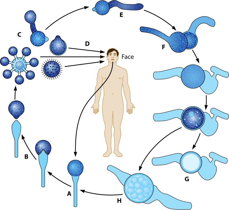
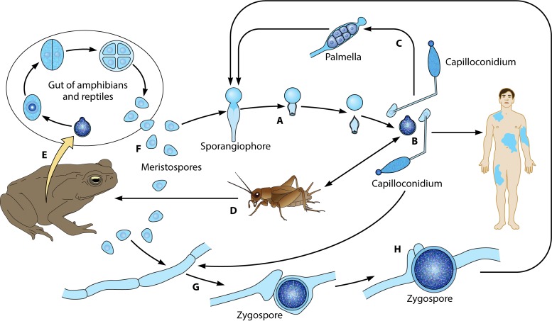
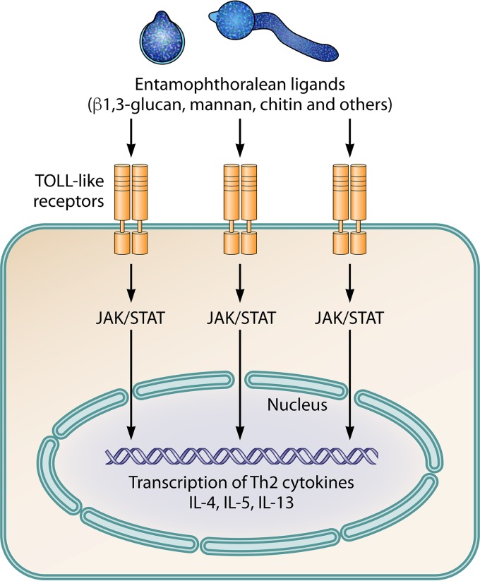
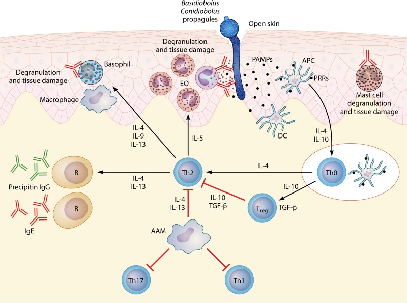
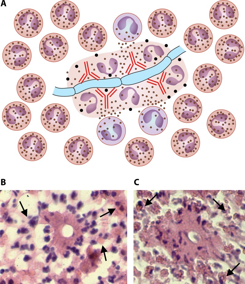
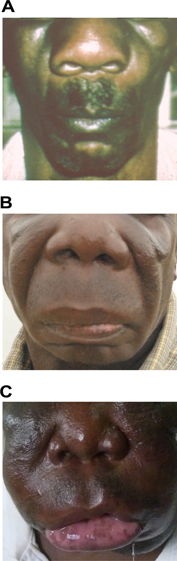
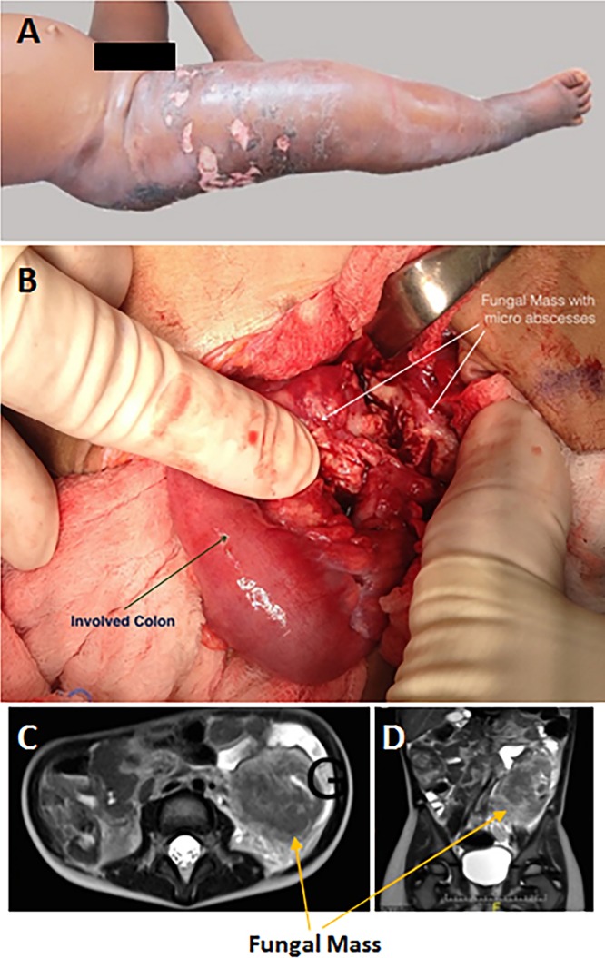
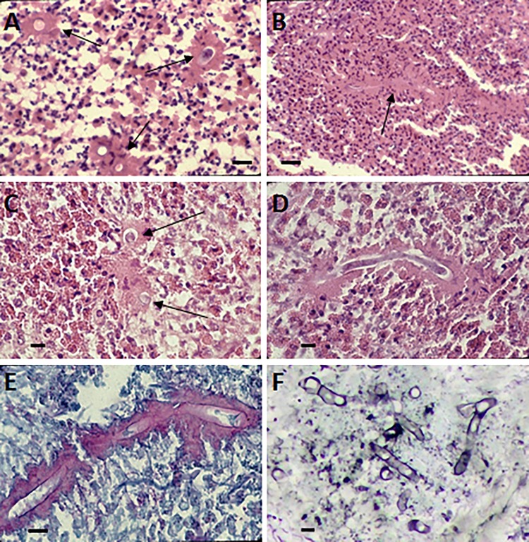
The pathogenic entomophthoralean fungi cause infection in insects and mammalian hosts. Basidiobolus and Conidiobolus species can be found in soil and insect, reptile, and amphibian droppings in tropical and subtropical areas. The life cycles of these fungi occur in these environments where infecting sticky conidia are developed. The infection is acquired by insect bite or contact with contaminated environments through open skin. Conidiobolus coronatus typically causes chronic rhinofacial disease in immunocompetent hosts, whereas some Conidiobolus species can be found in immunocompromised patients. Basidiobolus ranarum infection is restricted to subcutaneous tissues but may be involved in intestinal and disseminated infections. Its early diagnosis remains challenging due to clinical similarities to other intestinal diseases. Infected tissues characteristically display eosinophilic granulomas with the Splendore-Höeppli phenomenon. However, in immunocompromised patients, the above-mentioned inflammatory reaction is absent. Laboratory diagnosis includes wet mount, culture serological assays, and molecular methodologies. The management of entomophthoralean fungi relies on traditional antifungal therapies, such as potassium iodide (KI), amphotericin B, itraconazole, and ketoconazole, and surgery. These species are intrinsically resistant to some antifungals, prompting physicians to experiment with combinations of therapies. Research is needed to investigate the immunology of entomophthoralean fungi in infected hosts. The absence of an animal model and lack of funding severely limit research on these fungi.

Keywords: Basidiobolus; Conidiobolus; Entomophthorales; Entomophthoramycota; basidiobolomycosis; conidiobolomycosis; entomophthoramycosis; rhinoconidiobolomycosis; rhinoentomophthoramycosis.


Figures

FIG 1
FIG 1
(Aa) Formation of a sporangiophore of the genus Conidiobolus developing a sticky conidium on top of the structure. (b) As the conidium matures, an invagination is formed at the base of the conidium (arrows), exerting pressure on the base. (c) The invagination suddenly everts, propelling the conidium away from the sporangiophore. (Ba) Basidiobolus sporangiophore developing a conidium at the top of the structure. The subconidial portion of the sporangiophore accumulates liquid that becomes turgid (arrows). (b) The continuing accumulation of liquid exerts extreme pressure at the base, forming a subconidial vesicle, and the pressure results in rupture and the abrupt release of the conidium. (c) The subconidial structure remains attached, but it detaches upon landing.
FIG 2
FIG 2
(A) Yellow-white colony of Conidiobolus coronatus on 2% Sabouraud dextrose agar (SDA) from a primary culture after 48 h. The presence of a few aerial hyphae and satellite colonies due to the ejection of conidia from sporangiophores, detected on the lid of the SDA plate (data not shown), is also observed. (B to G) Formation of secondary conidia from a primary multinucleate conidium of C. coronatus in lactophenol blue. Bars, 20 μm (B, D, and G), 22 μm (C and E), and 21 μm (F). Note the different steps of development into fully formed corona secondary conidia in panels F and G. (H) Ejected secondary conidia and also several empty sporangiophores and three small ejected secondary conidia, some of which are developing germ tubes. Bar, 22 μm. (I and J) Single conidiophore with a secondary sporangiophore and conidium (I) and several primary C. coronatus conidia, one with several coenocytic hyphae (J). Bars, 20.0 μm (I) and 22 μm (J). (K and L) Presence of C. coronatus villose conidia in water agar cultures. Bars, 18 μm (K) and 19 μm (L).
FIG 3
FIG 3
(A) Powdery creamy colony of Conidiobolus incongruus at 72 h with a few aerial hyphae and some satellite colonies. (B and C) Sexual zygospores typical of C. incongruus in culture in lactophenol blue. Bars, 10 μm. (D) Multireplicative conidium bearing several secondary small conidia and a hyphal fragment (lactophenol blue). Bar, 8.0 μm. (E) Numerous primary conidia with sharp-pointed papillae. Some of the conidia are in the process of developing germ tubes, and at least one has a secondary replicative conidium. Bar, 15 μm.
FIG 4
FIG 4
(A) Creamy powdery colony of Conidiobolus lamprauges with radiating folds from the center of the colony at 72 h. (B and C) C. lamprauges smooth spherical sexual zygospores with thick cell walls in lactophenol blue. Bars, 20 μm (B) and 11 μm (C). (D and E) Asexual conidia found in culture plates of C. lamprauges (lactophenol blue). Note the smooth papilla projection of the conidia contrasting with that in C. incongruus (Fig. 3). Bars, 10 μm. (F) Secondary replicative conidium in lactophenol blue. Bar, 10 μm.
FIG 5
FIG 5
(A and B) The life cycle of Conidiobolus species starting with the development of a sporangiophore from hyphae (A) and the ejection of multireplicative primary conidia at the top of sporangiophores (B) (Fig. 2). (C and D) The primary conidia could replicate into secondary conidia that could also attach to passing hosts (C) or directly attach to the skin of humans (including the villose conidia of C. coronatus) (D). Clinical samples can be cultured, leading to the development of hyphae and sporangiophores (long arrow). (E to H) In nature and in culture, the primary conidia could form coenocytic filaments, which, after the interchange of genetic material, could lead to the formation of sexual zygospores, and the cycle starts all over again.
FIG 6
FIG 6
The life cycle of Basidiobolus ranarum. (A) The life cycle starts when sticky conidia are forcibly ejected from sporangiophores. (B) The sticky primary conidium could attach to a passing host (humans or insects) or develop an elongated adhesive conidium (capilloconidium), which also can attach to passing hosts. (C) The latter secondary elongated structure could develop to contain a sticky beak haptor that divides to form numerous “Palmella” endospores, some of which are released outside the broken capilloconidium cell wall, giving rise to new hyphae and single sporangiophores (A). (D and E) The target insects (D) can be ingested by reptiles or amphibians (E), initiating a new cycle inside the intestinal tract of these animals. (F) In this new environment, hundreds of resistant meristospores are produced and then secreted in feces. When environmental conditions are right, coenocytic hyphae are developed (G). (H) If two opposite-sex hyphae contact each other, their exchange of genetic material leads to the formation of sexual zygospores (see also Fig. 7). Zygospores can develop into sporangiophores (long arrow).
FIG 7
FIG 7
(A) Creamy rugose colony of Basidiobolus ranarum on 2% Sabouraud dextrose agar. (B to E) Encounter of opposite-sex hyphae before the formation of lateral beaks. Bars, 12 μm (B to D) and 10 μm (E). (F and G) After the exchange of genetic material, zygospores develop with their characteristic beak. Bars, 25 μm (F) and 15 μm (G).
FIG 8
FIG 8
Presentation of key antigenic molecules from invading entomophthoralean hyphae, based on the development of key Th2 cytokines and the typical eosinophilic reactions hypothetically triggered by entomophthoralean fungi during infection. The pathogen-associated molecular pattern (probably related to β-1,3-glucan, mannan, chitin, and others) could stimulate Toll-like cell receptors through signaling via Janus kinase (JAK) and signal transducer and activator of transcription (STAT). These molecules signal the nucleus to activate a cascade of events leading to the production of Th2-related cytokines (IL-4, IL-5, and IL-13), locking the host's immune system in a Th2 mode.
FIG 9
FIG 9
Putative events based on histopathological and immunological findings during entomophthoramycosis and during parasitic infections (112, 133, 137). Under this scenario, through open skin, a conidium attaches to the host and produces a germ tube penetrating the host. The invading hyphae then release secretory immunogens (pathogen-associated molecular patterns [PAMPs]). Dendritic cells (DC), through pattern recognition receptors (PRRs), contact the antigen and are activated, becoming antigen-presenting cells (APCs). The activated APCs process the antigens and release IL-4 during migration to nearby lymph nodes to present the antigen to Th0 naive cells. The Th0 naive cells in turn release IL-4 and IL-10 and become a powerful Th2 subset. The Th2 subset releases more IL-4, IL-5, IL-13, and IL-10, resulting in the downregulation of Th17 and Th1 subsets. These interleukins activate the differentiation of alternative activated macrophages (AAM) that could inhibit the proliferation of cells such as Th1, Th2, and Th17 cells. However, the exacerbated production of IL-4 and IL-5 by the Th2 subset drives the immune response into a strong Th2 subset. In turn, IL-4 and IL-13 stimulate B cells to produce precipitin IgG (detected by serological assays in cases of entomophthoramycosis) and IgE as well as the activation of effector cells such as mast cells, eosinophils (EO), and basophils. The released IgE will also specifically bind to the invading hyphae, and the eosinophils will in turn attach to the Fc region of IgE, triggering the degranulation of the eosinophils around the invading hyphae. A similar outcome occurs after IgE binding to mast cells and basophils, causing fibrosis and tissue damage, consistent with the clinical features of entomophthoramycosis. TGF-β, transforming growth factor β.
FIG 10
FIG 10
As highlighted in Fig. 9, the Th2 subset will trigger the release of IL-4, IL-5, and IL-13, and B cells will express IgE (red immunoglobulins). (A) The eosinophilic reaction around an invading hypha encases basidiobolomycosis or conidiobolomycosis. Note the presence of IgE, the degranulation of eosinophils around the hyphae, and several pyknotic nuclei within degranulate eosinophilic material. (B and C) Histopathological sections from a human case of intestinal basidiobolomycosis. Note the presence of numerous eosinophils surrounding the cross sections of hyphae that appear as spherical or oval structures in the center of the Splendore-Höeppli phenomenon. Under this perspective, the released IgE (red immunoglobulins) will bind to cell wall antigens that attract eosinophils to the site of infection. Panels A and B display the binding and degranulation of the eosinophils on the hyphae triggered by IgE (panel A, red immunoglobulins). The eosinophil nuclei at this stage appear at the periphery of the eosinophilic precipitate (A and B). As the lesion becomes old (chronic stages), other eosinophils will bind the complex, and after degranulation, their nuclei are also incorporated into the eosinophilic material and become pyknotic, giving rise to the Splendore-Höeppli phenomenon (C). In some instances, only the eosinophilic material will be expressed around the invading microbe (115, 137).
FIG 11
FIG 11
(A) An early case of conidiobolomycosis. (B and C) Two examples of late conidiobolomycosis infection. Extreme facial deformity of the patient is observed in panel B, whereas panel C depicts a case of facial elephantiasis caused by C. coronatus in a chronic nontreated case. (Panels B and C are reprinted from reference .)
FIG 12
FIG 12
(A) A 3-year-old girl with extensive swelling on the upper section of her right lower limb affecting the inguinal area and buttocks. The presence of multiple ulcers and skin discoloration is also evident. (Reprinted from reference with permission of the publisher.) (B) Exploratory intestinal laparotomy showing an edematous colon (arrow) surrounded by granulomatous masses displaying several microabscesses (arrows) in a case of human intestinal basidiobolomycosis. (C and D) Prior to surgery, MRI had shown a large predominantly hypointense retrocolic mass (arrows). (Panels B through D are reprinted from reference .)
FIG 13
FIG 13
(A) H&E-stained cross sections of Basidiobolus ranarum hyphae (arrows) from a case of intestinal basidiobolomycosis. Note the numerous eosinophils and the Splendore-Höeppli phenomenon. Bar, 20 μm. (B) Longitudinal B. ranarum hypha surrounded by numerous eosinophils and the Splendore-Höeppli phenomenon from a case of subcutaneous infection. Bar, 50 μm. (C and D) H&E-stained cross section (arrows) (bar, 10 μm) (C) and longitudinal hyphae of Conidiobolus coronatus surrounded by numerous eosinophils and the Splendore-Höeppli phenomenon (D) (bar, 12 μm) from a biopsy specimen of a conidiobolomycosis case involving the nostrils. (E) C. coronatus longitudinal hypha stained with periodic acid-Schiff stain. Bar, 10 μm. The content of the hypha is poorly stained, but the Splendore-Höeppli phenomenon is enhanced with the periodic acid-Schiff stain, indicating the presence of glycoproteins and polysaccharides. (F) C. coronatus stained with Grocott's methenamine silver. Bar, 8 μm. The presence of poorly stained coenocytic transverse and longitudinal filaments is evident.
None
None

References

    1. Ader P, Dodd JK. 1979. Mucormycosis and entomophthoromycosis. Mycopathologia 68:67–99. doi: 10.1007/BF00441088. - DOI - PubMed
    1. Blaché R, Destombes P, Nazimoff O. 1961. Mycoses sous-cutanées nouvelles an Sud-Cameroun. Bull Soc Path Exot 54:56–63.
    1. Bras G, Gordon CC, Emmons CW, Prendegast KM, Sugar M. 1965. A case of phycomycosis observed in Jamaica; infection with Entomphthora coronata. Am J Trop Med Hyg 14:141–145. doi: 10.4269/ajtmh.1965.14.141. - DOI - PubMed
    1. Brefeld O. 1884. Conidiobolus utricolosis und minor. Untersuchungen aus dem Gesamtgebiete der. Mykologie 4:75–78.
    1. Couch JN. 1939. A new Conidiobolus with sexual reproduction. Am J Bot 26:119–130. doi: 10.1002/j.1537-2197.1939.tb12878.x. - DOI

Substances

LinkOut - more resources