Review
. 2018 Aug 29;31(4):e00040-18.
doi: 10.1128/CMR.00040-18. Print 2018 Oct.

Nonsuppurative (Aseptic) Meningoencephalomyelitis Associated with Neurovirulent Astrovirus Infections in Humans and Animals

Affiliations
Review

Nonsuppurative (Aseptic) Meningoencephalomyelitis Associated with Neurovirulent Astrovirus Infections in Humans and Animals

Gábor Reuter et al. Clin Microbiol Rev. .

Abstract

Astroviruses are thought to be enteric pathogens. Since 2010, a certain group of astroviruses has increasingly been recognized, using up-to-date random amplification and high-throughput next-generation sequencing (NGS) methods, as potential neurovirulent (Ni) pathogens of severe central nervous system (CNS) infections, causing encephalitis, meningoencephalitis, and meningoencephalomyelitis. To date, neurovirulent astrovirus cases or epidemics have been reported for humans and domesticated mammals, including mink, bovines, ovines, and swine. This comprehensive review summarizes the virology, epidemiology, pathology, diagnosis, therapy, and future perspective related to neurovirulent astroviruses in humans and mammals, based on a total of 30 relevant articles available in PubMed (searched by use of the terms "astrovirus/encephalitis" and "astrovirus/meningitis" on 2 March 2018). A paradigm shift should be considered based on the increasing knowledge of the causality-effect association between neurotropic astroviruses and CNS infection, and attention should be drawn to the role of astroviruses in unknown CNS diseases.

Keywords: animal; astrovirus; encephalitis; human; meningitis; meningoencephalomyelitis; neurotropic; neurovirulent.


Figures

FIG 1
FIG 1
Representative sections from the anterior horn of the lumbar spinal cord for Ni-PoAstV-3-infected freshly weaned pigs with encephalomyelitis in stage 1 (posterior weakness) (A) and stage 3 (complete paralysis) (B) of the disease. The Ni-PoAstV-3-positive cells, which are presumed (based on the anatomical positions of the cells) to be motoneurons (arrows) and interneurons (arrowheads), were visualized using a digoxigenin-11-UTP (DIG)-labeled RNA probe and an in situ RNA hybridization technique. The 189-nt-long DIG-labeled antisense PoAstV-3 RNA probe targets the RdRp region of Ni-PoAstV-3 (accession numbers KY073229 to KY073232). The sections were visualized using an anti-DIG Fab fragment conjugated to alkaline phosphatase (Roche) and NBT/BCIP solution (Roche) supplemented with 1 mM levamisole. The section in panel A was counterstained with FastRed. Note that Ni-PoAStV-3 ISH-positive apoptotic (shrinking) neurons (double arrows in inserts) are observable in all stages of the disease.
FIG 2
FIG 2
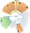
Phylogenetic analysis of full-length capsid proteins (ORF2) of representative members of the family Astroviridae. The amino acid sequences were aligned with the MUSCLE algorithm (86), and evolutionary analysis was conducted in MEGA 6 (87), using an ML method based on the LG model with frequencies, a discrete gamma distribution (+G), and evolutionarily invariable sites (+I). Bootstrap values were determined for 1,000 replicates. The Ni-AstV strains with known full-length capsid proteins are indicated with red squares. The astrovirus clades representing the known Ni-AstVs are highlighted in orange. The Avastroviruses (accession numbers AB033998, FR727149, HQ889774, FR727146, KJ020899, JF414802, Y15936, AF206663, EU143851, FJ434664, and FJ919228) were chosen as a tree outgroup. Abbreviations: BaAstV, bat astrovirus; BdAstV, bottlenose dolphin astrovirus; BoAstV, bovine astrovirus; CAstV, California sea lion astrovirus; CaAstV, canine astrovirus; ErAstV, European roller astrovirus; FeAstV, feline astrovirus; FoAstV, fox astrovirus; HAstV, human astrovirus; MiAstV, mink astrovirus; MuAstV, murine astrovirus; OvAstV, ovine astrovirus; PoAstV, porcine astrovirus; RaAstV, rabbit astrovirus; RAstV, rat astrovirus; WBAStV, wild boar astrovirus.
FIG 3
FIG 3
Representative genome map and identity graph (above the map) for Ni-AstVs. Vertical lines in the identity graph represent identical nucleotides (nt) in the alignment. Thicker lines represent multiple, consecutive identical nt. The identity graph was generated from the MUSCLE alignment of the complete genome/complete coding sequences of all known Ni-AstVs by use of UGENE ver 1.26 and CorelDraw ver. 12. The ruler above the identity graph represents nt positions in the alignment. The promoter sequence (gray background) of sgRNA is part of a conserved stem-loop (*). The secondary RNA structure of the sgRNA promoter was generated from the alignment of the corresponding genome region of all Ni-AstVs by use of the RNAalifold Web server (88) and VARNA visualization software, version 3.9. Note that s2m of the 3′ end is missing in the Ni-HAstV strains of MLB-1 (accession number LC064152) and MLB-2 (accession number KT224358). ORF, open reading frame; RFS, ribosomal frameshift signal; NSP-1a, nonstructural polyprotein translated from ORF1a; NSP-1ab, nonstructural polyprotein continuously translated from ORF1a and ORF1b; sgRNA, subgenomic RNA.
FIG 4
FIG 4
Schematic maps and identity graphs (above the maps) for nonstructural protein 1ab (NSP-1ab) and capsid for all known Ni-AstVs. Vertical lines in the identity graphs represent identical amino acids in the alignment. Thicker lines indicate multiple, consecutive identical aa. The identity graphs were generated from the MUSCLE alignments of NSP-1ab and capsid proteins of all known Ni-AstVs with complete genome sequences or complete CDS by use of UGENE ver 1.26 and CorelDraw ver. 12. The rulers above the identity graphs represent aa positions in the alignments. Hel, helicase; cc 1 to 3, coiled coils; TM 1 to 7, transmembrane domains; VPg, genome-associated viral protein; NLS, nuclear localization sequence; HVR, hypervariable region; RdRp, RNA-dependent RNA polymerase; RBD, RNA-binding domain. The red arrow shows a possible trypsin cleavage site, and yellow arrows show possible caspase cleavage sites.
FIG 5
FIG 5
Schematic representation of seven predicted transmembrane domains (yellow columns; TM1 to -7) identifiable in the N-terminal region of NSP-1ab of all known Ni-AstVs. Numbers in parentheses and italics indicate the first (numbers in the upper part) and last (numbers in the lower part) aa positions of the given TM domain. The predictions are based on the sequences listed on the left side of the figure and were determined using the MEMSAT software of the PSIPRED Protein Sequence Analysis Workbench (69, 70). All Ni-AstVs contain essentially the same number and topology of TM domains (data not shown).
FIG 6
FIG 6
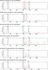
Representative probability (y axis) diagrams for predicted coiled-coil structures and their localizations in the aa sequences of NSP-1ab (x axis). The diagrams were generated using Ni-AstV reference sequences (indicated above the diagrams) and screening windows of 14, 21, and 28 aa (blue, green, and red lines) by use of the coiled-coil prediction software of ExPASy (71, 72). All other Ni-AstV sequences showed essentially the same localization of coiled coils (data not shown).
FIG 7
FIG 7
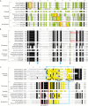
(A) Amino acid alignment of the N-terminal ends (RNA-binding domain [RBD]) of representative capsid sequences of neurovirulent (Ni) AstVs. Conservative and basic amino acids (R, K, and H) are shown with black and green backgrounds. Glutamine (Q) residues in Q-rich regions are marked with a deep yellow background and red boxes. Amino acid positions are shown according to the scale above the sequences. (B) Amino acid alignment and presumed trypsin cleavage sites (arrows) of the junctions of the conserved and hypervariable regions of representative neurovirulent (Ni) and “classical” AstV capsids. Red squares indicate N-terminal aa sequences of VP26 (HAstV2) and VP27/VP28 (HAstV8), experimentally identified by N-terminal sequencing (80, 89). Identical and highly conserved aa are marked with a black background in the alignment. Colored arrows indicate the conserved arginine (R) and lysine (K) residues which may act as potential trypsin cleavage sites (90), among which the conserved R/K residues marked with a green background and a red arrow are the most likely to be active. Numbers in parentheses indicate the first and last aa positions in the capsid polyprotein. (C) Representative amino acid alignment of the surroundings of the conserved acidic section (blue boxes), located in the C-terminal hypervariable regions, for all known neurovirulent (Ni; VA/HMO clade) and “classical” AstV capsids. Conservative and acidic amino acids (inside blue boxes) are shown with black and yellow backgrounds, respectively. Putative caspase cleavage sites of HAstV8 are marked in bold and underlined (81, 82). The conserved genome region that contains the Q(I/L)QxR(F/Y) motif, which is predominantly present among Ni-AstVs, is marked with a red box. Numbers in parentheses indicate the first and last aa positions in the capsid polyprotein.
None
None
None

References

    1. Glaser CA, Honarmand S, Anderson LJ, Schnurr DP, Forghani B, Cossen CK, Schuster FL, Christie LJ, Tureen JH. 2006. Beyond viruses: clinical profiles and etiologies associated with encephalitis. Clin Infect Dis 43:1565–1577. doi:10.1086/509330. - DOI - PubMed
    1. Long MT. Overview of meningitis, encephalitis, and encephalomyelitis. MSD veterinary manual. http://www.msdvetmanual.com/nervous-system/meningitis,-encephalitis,-and... Accessed 2 November 2017.
    1. World Organization for Animal Health. 2004. Manual of diagnostic tests and vaccines for terrestrial animals, 5th ed World Organization for Animal Health, Paris, France.
    1. Saylor D, Thakur K, Venkatesan A. 2015. Acute encephalitis in the immunocompromised individual. Curr Opin 28:330–336. doi:10.1097/YCO.0000000000000169. - DOI - PubMed
    1. Glaser CA, Gilliam S, Schnurr D, Forghani B, Honarmand S, Khetsuriani N, Fischer M, Cossen CK, Anderson LJ. 2003. In search of encephalitis etiologies: diagnostic challenges in the California Encephalitis Project, 1998–2000. Clin Infect Dis 36:731–742. doi:10.1086/367841. - DOI - PubMed

Publication types

MeSH terms

LinkOut - more resources