. 2018 Aug 29;10(9):463.
doi: 10.3390/v10090463.

The Virology of Taterapox Virus In Vitro

Affiliations

The Virology of Taterapox Virus In Vitro

Scott Parker et al. Viruses. .

Abstract

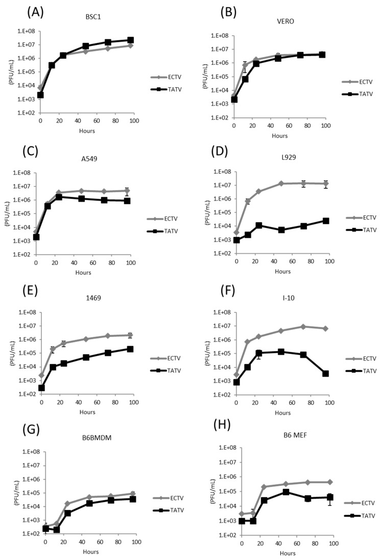
Taterapox virus (TATV) is phylogenetically the closest related virus to variola-the etiological agent of smallpox. Despite the similarity, few studies have evaluated the virus. In vivo, TATV can infect several animals but produces an inapparent infection in wild-type mice; however, TATV does cause morbidity and mortality in some immunocompromised strains. We employed in vitro techniques to compare TATV to ectromelia (ECTV) and vaccinia (VACV) viruses. Both ECTV and TATV replicate efficiently in primate cell lines but TATV replicates poorly in murine cells lines. Furthermore, TATV induces cytopathic effects, but to a lesser extent than ECTV, and changes cytoskeletal networks differently than both ECTV and VACV. Bioinformatic studies revealed differences in several immunomodulator open reading frames that could contribute to the reduced virulence of TATV, which were supported by in vitro cytokine assays.

Keywords: ectromelia; gerbil; orthopoxvirus; smallpox; taterapox; vaccinia; variola.


Conflict of interest statement

The authors declare no conflict of interest.

Figures

Figure 1
Figure 1
Alignment of the Complement Control Proteins from TATV, MPXV-ZAR, CPXV-GER91, VACV-COP, CMLV-CMS, VARV-BEN68, and ECTV-MOS. Regions of high consensus (90%) are shown in red; regions of low consensus (50%) are shown in blue.
Figure 2
Figure 2
ECTV and TATV growth kinetics. Cell monolayers of (A) BS-C-1, (B) Vero, (C) A549, (D) L929, (E) 1469, (F) I-10, (G) B6BMDM, and (H) B6 MEF cells were infected with a multiplicity of infection (MOI) of 0.01 PFU/cell and total virus production was measured by plaque-assays on BS-C-1 cells at various times up to 96 hours post-infection (h.p.i.). Representative data of three experiments.
Figure 3
Figure 3
VACV, ECTV, and TATV plaque morphologies on BS-C-1 cells stained at day three, five, and seven days post-infection (p.i). Data are representative of three experiments. 1× magnification.
Figure 4
Figure 4
BS-C-1 cells were infected with ECTV or TATV. Wells were overlaid with CMC to reveal standard plaques or were not overlaid with CMC to reveal comet-plaques. Data are representative of three experiments. 2× magnification.
Figure 5
Figure 5
Detection of microtubules and actin filaments by confocal microscopy. BS-C-1 cells were infected at an MOI of five (or mock, A) with either (B,E,H,K,N) VACV, (C,F,I,L,O) ECTV, or (D,G,J,M,P) TATV. After 4, 8, 12, 24, or 30 h.p.i., the cells were fixed, permeabilized, and stained. Nuclei (blue), F-actin (red), and microtubules (green) are shown. (A) Representative image of uninfected cells. (BD) Infected cells at four h.p.i.; (B) VACV cells are displaying the retracted, rounded CPE-M 1. (E) Migratory CPE-M 2 induced after VACV infection (H) at four h.p.i. Re-flattened, stretched CPE-M 3 observed at 12 h.p.i. with VACV. Inset depicts the virus-propelling actin tails displayed all over the infected cell body. (K) Pre-terminal stage of VACV infection. The CPE-M 4 represents the onset of cell death. Inset depicts the formation of projections at the cell periphery. (N) End-stage of VACV infection is observed here and represented by CPE-M 5. The infected cell displays a multi-branched morphology, with several protrusions formed by both actin and microtubule filaments. This is the terminal stage of VACV infection and CPE-M 5 evolves to cell death. TATV and ECTV cells are shown for comparison. 1000x magnification.
Figure 6
Figure 6
The occurrence of CPE morphotypes one to five in VACV-, ECTV-, and TATV-infected BS-C-1 cells. Cells were infected and prepared as outlined in the legend of Figure 5. The percentage of cells with (AE) morphotype 1–5, respectively, were reported at 4, 8, 12, 24, and 30 h.p.i.
Figure 7
Figure 7
BS-C-1 cells were infected at an MOI of five, as described previously. After 4, 8, 12, 24, and 30 h.p.i., cells were fixed and stained with Rhodamine-conjugated Phalloidin to mark the actin cytoskeleton, as well as with the anti-VACV B5 envelope protein. B5-positively marked actin tail accumulation over the course of infection with the different OPVs, VACV, TATV, or ECTV, was quantified and plotted. (A) The average number of actin tails per cell during infection. (B) The percentage of cells displaying at least one B5-positve actin tail in the cell body was determined during the time-course of infection with VACV, TATV, or ECTV. Graphs were plotted with Graphpad Prism 6.0. Data are representative of three experimental replications.

References

    1. Chen N., Bellone C.J., Schriewer J., Owens G., Fredrickson T., Parker S., Buller R.M. Poxvirus interleukin-4 expression overcomes inherent resistance and vaccine-induced immunity: Pathogenesis, prophylaxis, and antiviral therapy. Virology. 2011;409:328–337. doi: 10.1016/j.virol.2010.10.021. - DOI - PMC - PubMed
    1. Esposito J.J., Knight J.C. Orthopoxvirus DNA: A comparison of restriction profiles and maps. Virology. 1985;143:230–251. doi: 10.1016/0042-6822(85)90111-4. - DOI - PubMed
    1. Esposito J.J., Sammons S.A., Frace A.M., Osborne J.D., Olsen-Rasmussen M., Zhang M., Govil D., Damon I.K., Kline R., Laker M., et al. Genome sequence diversity and clues to the evolution of variola (smallpox) virus. Science. 2006;313:807–812. doi: 10.1126/science.1125134. - DOI - PubMed
    1. Lourie B., Nakano J.H., Kemp G.E., Setzer H.W. Isolation of poxvirus from an African Rodent. J. Infect. Dis. 1975;132:677–681. doi: 10.1093/infdis/132.6.677. - DOI - PubMed
    1. Parker S., Crump R., Hartzler H., Buller R.M. Evaluation of Taterapox Virus in Small Animals. Viruses. 2017;9:203. doi: 10.3390/v9080203. - DOI - PMC - PubMed

Publication types

MeSH terms

LinkOut - more resources