. 2018 Aug 29;9(1):3501.
doi: 10.1038/s41467-018-05742-z.

In vivo phosphoproteomics reveals kinase activity profiles that predict treatment outcome in triple-negative breast cancer

Affiliations

In vivo phosphoproteomics reveals kinase activity profiles that predict treatment outcome in triple-negative breast cancer

Ivana Zagorac et al. Nat Commun. .

Abstract

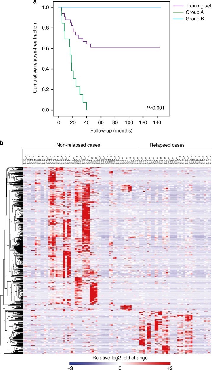
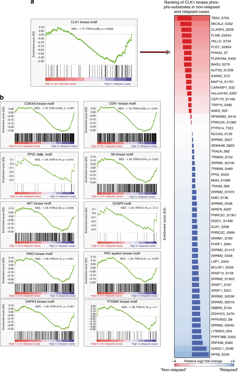
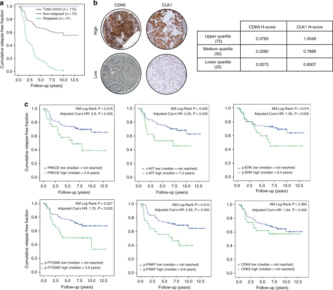
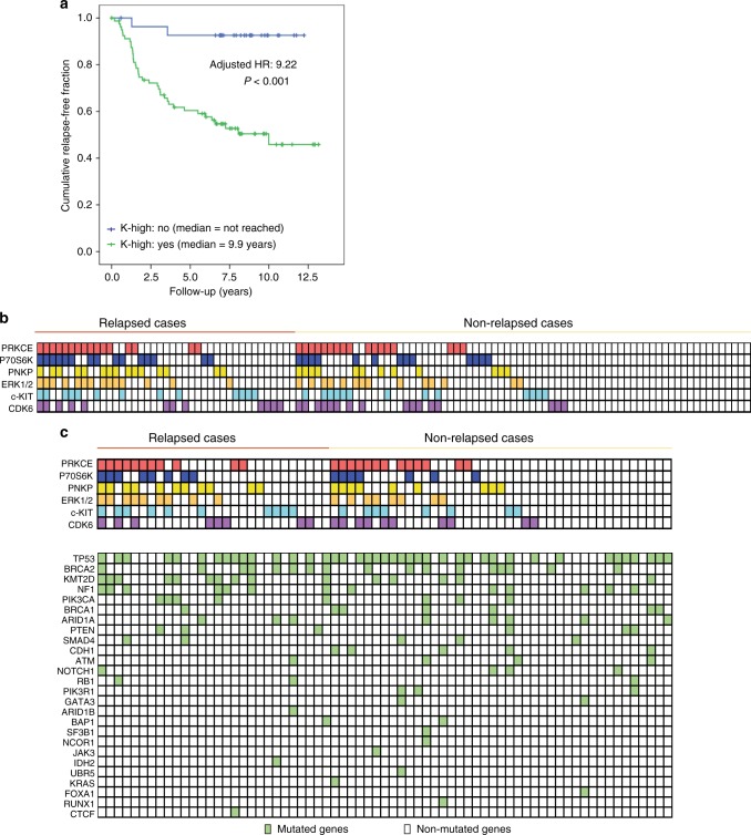
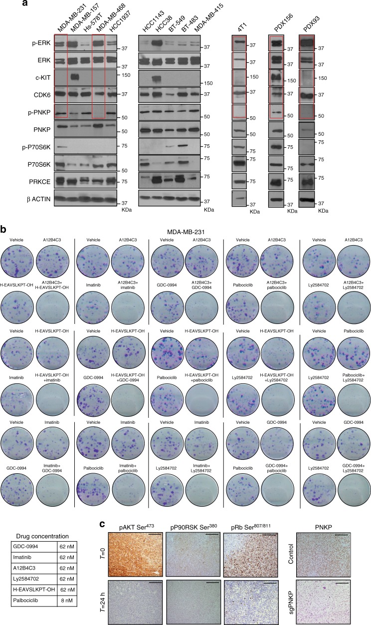
Triple-negative breast cancer (TNBC) lacks prognostic and predictive markers. Here, we use high-throughput phosphoproteomics to build a functional TNBC taxonomy. A cluster of 159 phosphosites is upregulated in relapsed cases of a training set (n = 34 patients), with 11 hyperactive kinases accounting for this phosphoprofile. A mass-spectrometry-to-immunohistochemistry translation step, assessing 2 independent validation sets, reveals 6 kinases with preserved independent prognostic value. The kinases split the validation set into two patterns: one without hyperactive kinases being associated with a >90% relapse-free rate, and the other one showing ≥1 hyperactive kinase and being associated with an up to 9.5-fold higher relapse risk. Each kinase pattern encompasses different mutational patterns, simplifying mutation-based taxonomy. Drug regimens designed based on these 6 kinases show promising antitumour activity in TNBC cell lines and patient-derived xenografts. In summary, the present study elucidates phosphosites and kinases implicated in TNBC and suggests a target-based clinical classification system for TNBC.


Conflict of interest statement

The authors declare no competing interests.

Figures

Fig. 1
Fig. 1
Phosphoprofiling of the patient training set. a There were two patient subgroups in the training set: A 13 patients who relapsed within <3 years after locoregional treatment (green chart), and B 21 patients who did not relapse >10 years after locoregional treatment (blue chart). The graphic shows the Kaplan–Meier curves of the training set as a whole (purple chart) and of the two subgroups. Median time to relapse in group A: 17.4 months; median time to relapse in group B: not reached (log-rank test, p < 0.001). b The heatmap shows the phosphopeptides (n = 702: 543 upregulated in non-relapsed and 159 upregulated in relapsed patients) with significant differences in phosphorylation levels in tumors from the training set by relapse status
Fig. 2
Fig. 2
Kinases driving the profiles of the relapsed cases. a Example of a chart of normalized enrichment scores (NESs) (left) obtained for CLK1 from the relative abundance of its phosphorylated substrates in either the relapsed or the non-relapsed cases (right)—or kinase set enrichment analysis (KSEAS). Each substrate (phosphopeptide) is represented in the KSEA as a vertical black line. The proteins to which they map are represented in the right column by their encoding genes, adjacent to the site at which phosphorylation was detected. In this column, a larger or shorter horizontal bar depicts, for each substrate, the Log2-fold regulation in the relapsed (blue) versus non-relapsed (red) cases. b Two phosphatase (DUSP6 and PP2C-δ) and 9 kinase domains were enriched in the relapsed cases. The finding of an enriched phosphatase domain can be accounted for by the presence of a high concentration of a substrate for that phosphatase in a specific subgroup of patient tumors or cell lines. The in silico tool cannot predict whether a phosphatase is functional based on the absence of phosphorylation of its putative substrates; however, it can predict which upstream kinases or phosphatases can bind (and phosphorylate or cleave) an identified substrate. P-values and false discovery rates (FDR) are depicted for each kinase or phosphatase. Although most KSEAS show a low FDR, a relaxed FDR boundary (up to 0.25) was allowed to ensure as little information loss as possible in the mass spectrometry-to-immunohistochemistry translation step
Fig. 3
Fig. 3
Independent validation. a Kaplan–Meier survival curve of the 113 patients comprising the first independent validation set—Val-1 (black chart); 72 of these patients did not relapse during 12.5 + years of observation (blue chart), whereas 41 patients did relapse (green chart). b Immunohistochemically stained examples of CDK6 and CLK1 from patients with H-scores in the upper (high) or lower (low) quartiles. c Kaplan–Meier curves according to the status (H-scores in the upper quartiles [green charts] or lower quartiles [blue charts]) of PRKCE, c-Kit, pERK (Thr202/Tyr204), pP70S6K (Thr389), pPNKP (Ser114/Thr118), and CDK6. The upper p-values shown in each chart were derived from the log-rank test comparing median overall relapse-free survival times (KM Log-Rank p value) between patients in the upper quartiles versus the lower quartiles. The hazard ratios depicted below correspond to those for relapses attributed to each kinase in a Cox model adjusted by T, N, G, and age, and are all statistically significant (Cox P< 0.005)
Fig. 4
Fig. 4
Clinical implications of kinomic and genomic landscapes. a Prognostic impact of the presence of one or more activated kinases: Kaplan–Meier curve for relapse of patients in validation set Val-1, whose tumors showed high activity of any of the 6 kinases (green chart: K-high = yes) versus the remaining (patients who showed low activity of the 6 kinases; blue chart: K-high = no). b Each of the 6 kinases with prognostic power is listed on the left side (rows), and for each patient (columns, stratified according to relapse status), the number of kinases with high-quartile staining are shown. c Below the kinase grid, a second grid contains the mutational status (yes/no, according to the filters explained in the Methods section) of the 25 most frequently mutated genes for each sequenced patient. The data show the “collapse” of mutational patterns into kinomic patterns; for instance, pattern 38 (Supplementary Table 5) was observed in patients both with and without TP53, BRCA2, PTEN, or ARID1A mutations. The same statement was also valid for other frequently observed kinase patterns in the validation set, such as c-Kit-high (pattern 36 (N = 9 patients), which accounted for patients with various combinations of wild-type and mutant TP53, BRCA2, PIK3CA, and PTEN) or CDK6-high
Fig. 5
Fig. 5
In vitro therapeutic efficacy and in vivo pharmacodynamics. a Western blotting showing the levels of each of the 6 kinases in the signature, in addition to their non-phosphorylated controls where applicable, in 10 human TNBC cell lines, the transplantable murine TNBC tumor model 4T1, and 2 patient-derived xenografts (PDXs). The three targets against which clinical-grade drugs are available are highlighted in red. b Colony assays (MDA-MB-231) showing the differences between single-target versus two-target pharmacological blockade. For each of the 15 possible 2-by-2 drug combinations using the 6 agents against the kinases in K-high, a representative vehicle-treated well, representative single-agent-treated wells and a well containing the doublet are shown. All 4-well images belong to unique 12-well dishes. Representative images of three independent experiments. c In vivo dosage of imatinib, GDC-0994, and palbociclib at standard doses for animal use led to decreased AKT, P90RSK, and RB phosphorylation levels (targets of the kinases c-Kit, ERK, and CDK6, respectively) in MDA-MB-231-xenografted tumors after 24 h. The right panel shows total PNKP levels in wild-type (upper) and CRISPR PNKP MDA-MB-231 transfectant (lower panels) tumors. Scale bar, 50 μm
Fig. 6
Fig. 6
In vivo therapeutic efficacy. a MDA-MB-231 is a cell line with high levels of p-ERK and CDK6, but no visible levels of c-Kit. It can be observed that, although imatinib-based doublets significantly prolonged mice overall survival as compared with the singlets or vehicle, the magnitude of the improvement is much lower than that seen with the p-ERK + CDK6-targeting doublet (GDC-0994 and palbociclib, mid chart; >5-fold increase in overall survival compared with ~2-fold). Below the Kaplan-Meier curves, the median overall survival (days) for each combination (or single-agent) in addition to the Log-Rank P-values are shown. Finally, representative tumor burdens of each treatment group are depicted. b MDA-MB-231 shows relatively high p-PNKP levels; CRISPR PNKP MDA-MB-231 transfectant xenografts treated with GDC-0994 also showed statistically significantly longer overall survival as compared with GDC-0994 administered to wild-type MDA-MB-231 or untreated CRISPR PNKP transfectants. Mice treated with: vehicle (n = 5), imatinib (n = 6), GDC-0994 (n = 4), palbociclib (n = 5), GDC-0994 + palbociclib (n = 5), palbociclib + imatinib (n = 5), sgPNKP (n = 11) and sgPNKP + GDC-0994 (n = 11). c Compared to MDA-MB-231, the levels of p-PNKP in MDA-MB-468 are almost undetectable. The levels of p-ERK and CDK6 are high, and those of c-Kit are low. Matching the observations in the other models, when a targeted doublet includes a target with low or absent expression (namely, p-PNKP in MDA-MB-468), no synergy is observed. In the three Kaplan-Meier curves it can be observed than the doublet is not better than any of the monotherapies (or sgPNKP alone). Representative tumor burden charts are shown below the survival curves. Mice treated with: vehicle (n = 6), sgPNKP and sgPNKP + imatinib (n = 5), imatinib (n = 8), GDC-0994 (n = 11), GDC-0994 + imatinib (n = 13), palbociclib (n = 12), GDC-0994 + palbociclib (n = 9), sgPNKP + palbociclib (n = 4) and sgPNKP + GDC-0994 (n = 7). d Finally, in PDX156 (c-Kit and pERK higher than CDK6), imatinib plus GDC-0994, and GDC-0994 plus palbociclib significantly prolonged median overall survival compared to the monotherapies. Mice treated with: vehicle (n = 5), imatinib (n = 4), GDC-0994 (n = 4), palbociclib (n = 4), GDC-0994 + palbociclib (n = 5) and GDC-0994 + imatinib (n = 9). In tumor burden graph, each point represents a tumor. The data are represented as mean±SEM and Student ´s t test was performed. *p < 0.05, **p < 0.01, ***p < 0.001

References

    1. Foulkes WD, Smith IE, Reis-Filho JS. Triple-negative breast cancer. N. Engl. J. Med. 2010;363:1938–1948. doi: 10.1056/NEJMra1001389. - DOI - PubMed
    1. Lehmann BD, et al. Identification of human triple-negative breast cancer subtypes and preclinical models for selection of targeted therapies. J. Clin. Invest. 2011;121:2750–2767. doi: 10.1172/JCI45014. - DOI - PMC - PubMed
    1. Curtis C, et al. The genomic and transcriptomic architecture of 2,000 breast tumours reveals novel subgroups. Nature. 2012;486:346–352. doi: 10.1038/nature10983. - DOI - PMC - PubMed
    1. Shah SP, et al. The clonal and mutational evolution spectrum of primary triple-negative breast cancers. Nature. 2012;486:395–399. doi: 10.1038/nature10933. - DOI - PMC - PubMed
    1. Banerji S, et al. Sequence analysis of mutations and translocations across breast cancer subtypes. Nature. 2012;486:405–409. doi: 10.1038/nature11154. - DOI - PMC - PubMed

Publication types

LinkOut - more resources