Review
. 2018 May-Aug;22(2):279.
doi: 10.4103/jomfp.JOMFP_125_15.

A review of artifacts in histopathology

Affiliations
Review

A review of artifacts in histopathology

Syed Ahmed Taqi et al. J Oral Maxillofac Pathol. 2018 May-Aug.

Abstract

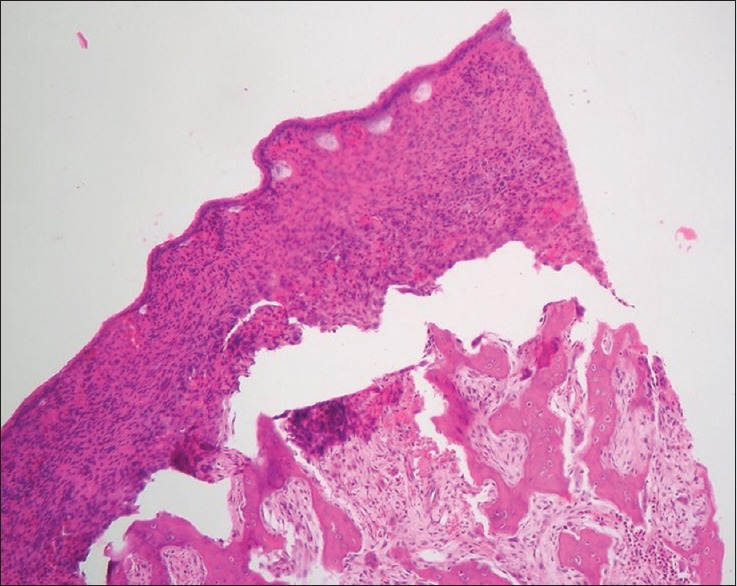
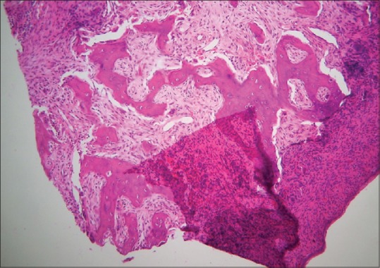
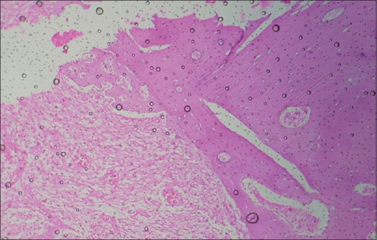
Histopathological examination is considered as gold standard procedure for arriving at a final diagnosis of various lesions of the human body. However, it is limited by a number of alterations of normal morphologic and cytological features that occur as a result of presence of artifacts. These artifacts may occur during surgical removal, fixation, tissue processing, embedding and microtomy and staining and mounting procedures. They can even lead to complete uselessness of the tissue. It is therefore essential to identify the commonly occurring artifacts during histopathological interpretations of tissue sections. This article reviews the common artifacts encountered during slide examination alongside the remedial measures which can be undertaken to differentiate between an artifact and tissue constituent.

Keywords: Artifact; diagnosis; histopathology; microtome; tissue specimen.


Conflict of interest statement

There are no conflicts of interest.

Figures

Figure 1
Figure 1
Histopathological image shows separation of connective tissue bands due to intralesional injection of anesthetic solution (H&E stain, ×10)
Figure 2
Figure 2
Histopathological image shows hemorrhage occurring during surgery which may be misinterpreted as pathological change (H&E, ×10)
Figure 3
Figure 3
Histopathological image shows tissue tear due to rough handling by forcep (H&E, ×10)
Figure 4
Figure 4
Histopathological image shows tissue tear and folds due to rough handling by forcep (H&E, ×10)
Figure 5
Figure 5
Histopathological image shows crush artifact (H&E, ×10)
Figure 6
Figure 6
Histopathological image shows sutural artifact (H&E, ×10)
Figure 7
Figure 7
Histopathological image shows tissue autolysis due to delayed fixation (H&E, ×10)
Figure 8
Figure 8
Histopathological image shows separation of epithelium from the connective tissue simulating vesiculobullous lesion (H&E, ×10)
Figure 9
Figure 9
Histopathological image shows bone trabeculae stained strongly with hematoxylin due to incomplete decalcification (H&E, ×10)
Figure 10
Figure 10
Histopathological image shows cholesterol clefts in odontogenic cysts (H&E, ×10)
Figure 11
Figure 11
Histopathological image shows scoring and tearing of section due to nick in knife edge (H&E, ×10)
Figure 12
Figure 12
Histopathological image shows venetian blind artifact due to vibration of knife edge (H&E, ×10)
Figure 13
Figure 13
Histopathological image shows displacement of bone during microtomy in association with the use of dull knife (H&E, ×10)
Figure 14
Figure 14
Histopathological image shows tangential cut artifact (H&E, ×10)
Figure 15
Figure 15
Histopathological image shows curling artifact due to folding of tissue due to blunt microtome knife (H&E, ×10)
Figure 16
Figure 16
Histopathological image shows wrinkles and folds due to uneven stretching of tissue sections (H&E, ×10)
Figure 17
Figure 17
Histopathological image shows stain deposit due to formation of fluorescent sheen in hematoxylin solution (H&E, ×10)
Figure 18
Figure 18
Histopathological image shows air bubbles formed during mounting procedure (H&E, ×10)

References

    1. Seoane J, Varela-Centelles PI, Ramírez JR, Cameselle-Teijeiro J, Romero MA. Artefacts in oral incisional biopsies in general dental practice: A pathology audit. Oral Dis. 2004;10:113–7. - PubMed
    1. Rastogi V, Puri N, Arora S, Kaur G, Yadav L, Sharma R, et al. Artefacts: A diagnostic dilemma – A review. J Clin Diagn Res. 2013;7:2408–13. - PMC - PubMed
    1. Margarone JE, Natiella JR, Vaughan CD. Artifacts in oral biopsy specimens. J Oral Maxillofac Surg. 1985;43:163–72. - PubMed
    1. Ficarra G, McClintock B, Hansen LS. Artefacts created during oral biopsy procedures. J Craniomaxillofac Surg. 1987;15:34–7. - PubMed
    1. Oliver RJ, Sloan P, Pemberton MN. Oral biopsies: Methods and applications. Br Dent J. 2004;196:329–33. - PubMed