Review
. 2018 Aug 12:2018:5697846.
doi: 10.1155/2018/5697846. eCollection 2018.

Radiological Patterns of Lung Involvement in Inflammatory Bowel Disease

Affiliations
Review

Radiological Patterns of Lung Involvement in Inflammatory Bowel Disease

Diletta Cozzi et al. Gastroenterol Res Pract. .

Abstract

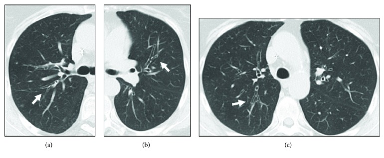
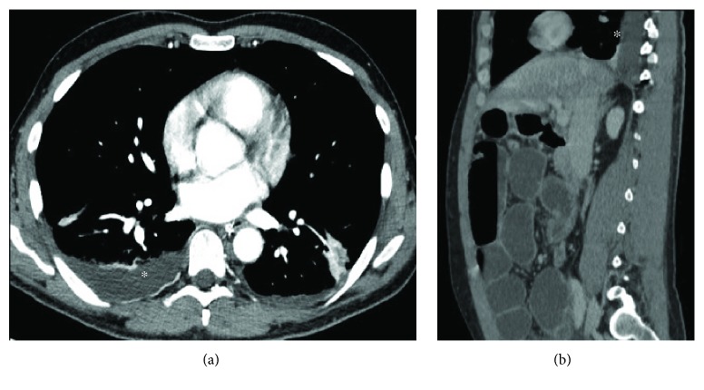
Inflammatory bowel disease (IBD) is a form of chronic inflammation of the gastrointestinal tract, including two major entities: ulcerative colitis and Crohn's disease. Although intestinal imaging of IBD is well known, imaging of extraintestinal manifestations is not extensively covered. In particular, the spectrum of IBD-associated or related changes in the chest is broad and may mimic other conditions. The common embryonic origin of intestine and lungs from the foregut, autoimmunity, smoking, and bacterial translocation from the colon may all be involved in the pathogenesis of these manifestations in IBD patients. Chest involvement in IBD can present concomitant with or years after the onset of the bowel disease even postcolectomy and can affect more than one thoracic structure. The purpose of the present paper is to present the different radiological spectrum of IBD-related chest manifestations, including lung parenchyma, airways, serosal surfaces, and pulmonary vasculature. The most prevalent and distinctive pattern of respiratory involvement is large airway inflammation, followed by lung alterations. Pulmonary manifestations are mainly detected by pulmonary function tests and high-resolution computed tomography (HRCT). It is desirable that radiologists know the various radiological patterns of possible respiratory involvement in such patients, especially at HRCT. It is essential for radiologists to work in multidisciplinary teams in order to establish the correct diagnosis and treatment, which rests on corticosteroids at variance with any other form of bronchiectasis.


Figures

Figure 1
Figure 1
The flowchart indicates when to perform chest X-ray and HRCT in IBD patients.
Figure 2
Figure 2
A 77-year-old woman with old history of CD. These HRCT coronal (a) and axial (b) reconstructions show a small nodule in the tracheal wall, most likely referring to a granuloma (arrows).
Figure 3
Figure 3
A 55-year-old woman with CD. Chest X-ray shows right basal bronchial wall thickening suggesting bronchitis (a). Axial HRCT study shows peribronchial wall thickening associated with bronchiolectasis (arrow) and signs of subpleural emphysema (b).
Figure 4
Figure 4
Axial HRCT scan (a) and sagittal reconstruction (b) show bilateral bronchiectasis (arrows) and some small mucoid impactions (blue arrowhead).
Figure 5
Figure 5
Multiple bilateral bronchiectasis in a 72-year-old woman with CD (arrows). Axial HRCT scans (a, b, c).
Figure 6
Figure 6
HRCT sagittal reconstruction. Cylindrical and varicose bronchiectasis (arrow) in the middle lobe in a 62-year-old woman with CD. A mosaic pattern due to air trapping is present.
Figure 7
Figure 7
Sagittal HRCT of a 52-year-old male patient with CD shows lower bronchiectasis and bronchiolectasis, associated with diffuse basal parenchymal opacities (ground glass pattern) and interstitial septal thickening.
Figure 8
Figure 8
Reverse halo or atoll sign in a patient with UC and possible OP.
Figure 9
Figure 9
A 67-year-old woman with CD. In both basal posterior segments of the lower lobes, an interlobular-intralobular interstitial thickening associated with ground glass opacities and some traction bronchiectasis and bronchiolectasis is appreciable. A subpleural sparing is associated. These findings are compatible with NSIP. Axial HRCT study (a) and sagittal reconstruction (b).
Figure 10
Figure 10
A 36-year-old woman with UC and previous total colectomy. In the basal posterolateral segment of lower lobes (more evident in the left), a fine interstitial thickening associated with ground glass opacities and some traction bronchiolectasis is appreciable. A subpleural sparing is associated (arrow). These findings are consistent with NSIP. Axial HRCT study (a) and sagittal reconstruction (b).
Figure 11
Figure 11
A 55-year-old woman with coexisting CD and GPA disease. These HRCT images (axial (a), coronal (b), sagittal (c)) show a pulmonary necrotic nodule in the lower left lobe: it is possible to notice the necrotic cavity inside. Whether this is due to CD of GPA was unclear.
Figure 12
Figure 12
An 18-year-old male with Crohn's disease in the acute phase and no additional comorbidities, who presented with mild shortness of breath. Coronal and axial reconstruction of contrast-enhanced chest CT reveals bilateral pulmonary embolism in both main pulmonary arteries (arrows, a-b). Coronal reconstruction for lung parenchyma (c).
Figure 13
Figure 13
A 43-year-old man with Crohn's disease subjected to previous surgical ileal resection. Asterisks () show unilateral pleural effusion during acute abdomen due to intestinal obstruction. Axial CT study (a) and sagittal reconstruction (b).

References

    1. Baumgart D. C., Carding S. R. Inflammatory bowel disease: cause and immunobiology. The Lancet. 2007;369(9573):1627–1640. doi: 10.1016/S0140-6736(07)60750-8. - DOI - PubMed
    1. Rodriguez-Roisin R., Bartolome S. D., Huchon G., Krowka M. J. Inflammatory bowel diseases, chronic liver diseases and the lung. European Respiratory Journal. 2016;47(2):638–650. doi: 10.1183/13993003.00647-2015. - DOI - PubMed
    1. Danese S., Fiocchi C. Ulcerative colitis. The New England Journal of Medicine. 2011;365(18):1713–1725. doi: 10.1056/NEJMra1102942. - DOI - PubMed
    1. Loftus E. V., Jr. Clinical epidemiology of inflammatory bowel disease: incidence, prevalence and environmental influences. Gastroenterology. 2004;126(6):1504–1517. doi: 10.1053/j.gastro.2004.01.063. - DOI - PubMed
    1. Papanikolaou I., Kagouridis K., Papiris S. A. Patterns of airway involvement in inflammatory bowel diseases. World Journal of Gastrointestinal Pathophysiology. 2014;5(4):560–569. doi: 10.4291/wjgp.v5.i4.560. - DOI - PMC - PubMed

LinkOut - more resources