

In Vivo Comparative Evaluation of Periapical Healing in Response to a Calcium Silicate and Calcium Hydroxide Based Endodontic Sealers
- PMID: 30159080
- PMCID: PMC6108798
- DOI: 10.3889/oamjms.2018.293
In Vivo Comparative Evaluation of Periapical Healing in Response to a Calcium Silicate and Calcium Hydroxide Based Endodontic Sealers
Abstract
Background: The composition of the root canal filling materials together with the apical limit of the root canal obturation affect the complete periapical healing after root canal therapy.
Aim: This study was performed to evaluate and compare the periapical healing in response to calcium-silicate (iRoot SP) and calcium-hydroxide (Apexit) based-sealers.
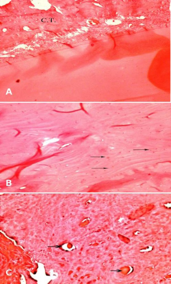
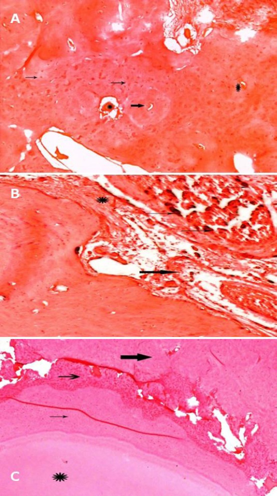
Material and methods: Seventy-two upper premolars root canals of six dogs were used. The teeth were randomly assigned to four groups: Group one: roots were obturated using gutta-percha and Apexit-sealer; Group two: roots were obturated using gutta-percha&iRoot SP-sealer; Group three: the teeth were left open without obturation; Group four: where healthy teeth were used as a negative control. Teeth were evaluated after one, two and three months. The newly formed mineralised apical tissue and the periapical inflammatory infiltrate of the obtained photomicrographs were evaluated, and scorings were statistically-analysed.
Results: The mean percentage of the periapical inflammatory infiltrates and mineralisation scoring after one, two and three months evaluation period were not significantly different among the four groups (P > 0.05).
Conclusions: Regardless of the sealer used, iRoot SP and Apexit promote healing of periapical tissues. IRoot SP sealer showed early insignificant more partial and almost full healing after two and three months.
Keywords: Bioceramic sealer; Calcium hydroxide; Root canal sealer.
Figures
References
-
- Wu MK, Tigos E, Wesselink PR. An l8-month longitudinal study on a new silicon-based sealer, RSA RoekoSeal:A leakage study in vitro. Oral Surgery, Oral Medicine, Oral Pathology, Oral Radiology, and Endodontology. 2002;94(4):499–502. https://doi.org/10.1067/moe.2002.124859. - PubMed
-
- Schäfer E, Zandbiglari T. Solubility of root?canal sealers in water and artificial saliva. International endodontic journal. 2003;36(10):660–9. https://doi.org/10.1046/j.1365-2591.2003.00705.x PMid:14511222. - PubMed
-
- Leonardo MR, Utrilla LS, Assed S, Ether SS. Calcium hydroxide root canal Sealers—Histopathologic evaluation of apical and peripaical repair after endodontic treatment. Journal of endodontics. 1997;23(7):428–32. https://doi.org/10.1016/S0099-2399(97)80296-8. - PubMed
-
- Al-Awadhi S, Spears R, Gutmann JL, Opperman LA. Cultured primary osteoblast viability and apoptosis in the presence of root canal sealers. J Endod. 2004;30:527–33. https://doi.org/10.1097/00004770-200407000-00016 PMid:15220652. - PubMed
LinkOut - more resources
Full Text Sources
Other Literature Sources