. 2018 Aug 7:2018:4971261.
doi: 10.1155/2018/4971261. eCollection 2018.

Exosomes Derived from miR-214-Enriched Bone Marrow-Derived Mesenchymal Stem Cells Regulate Oxidative Damage in Cardiac Stem Cells by Targeting CaMKII

Affiliations

Exosomes Derived from miR-214-Enriched Bone Marrow-Derived Mesenchymal Stem Cells Regulate Oxidative Damage in Cardiac Stem Cells by Targeting CaMKII

Yan Wang et al. Oxid Med Cell Longev. .

Abstract

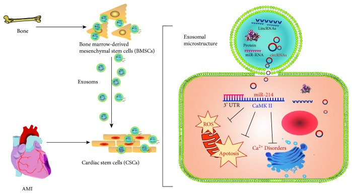
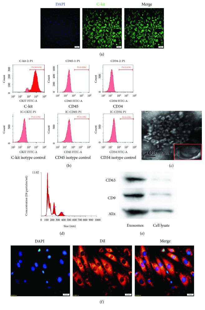
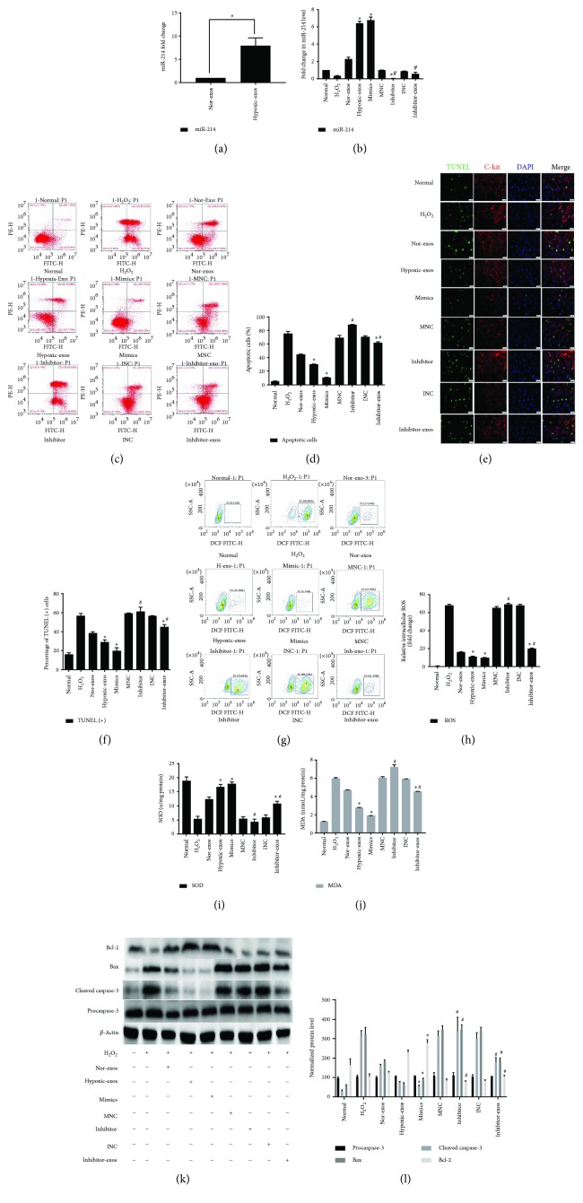
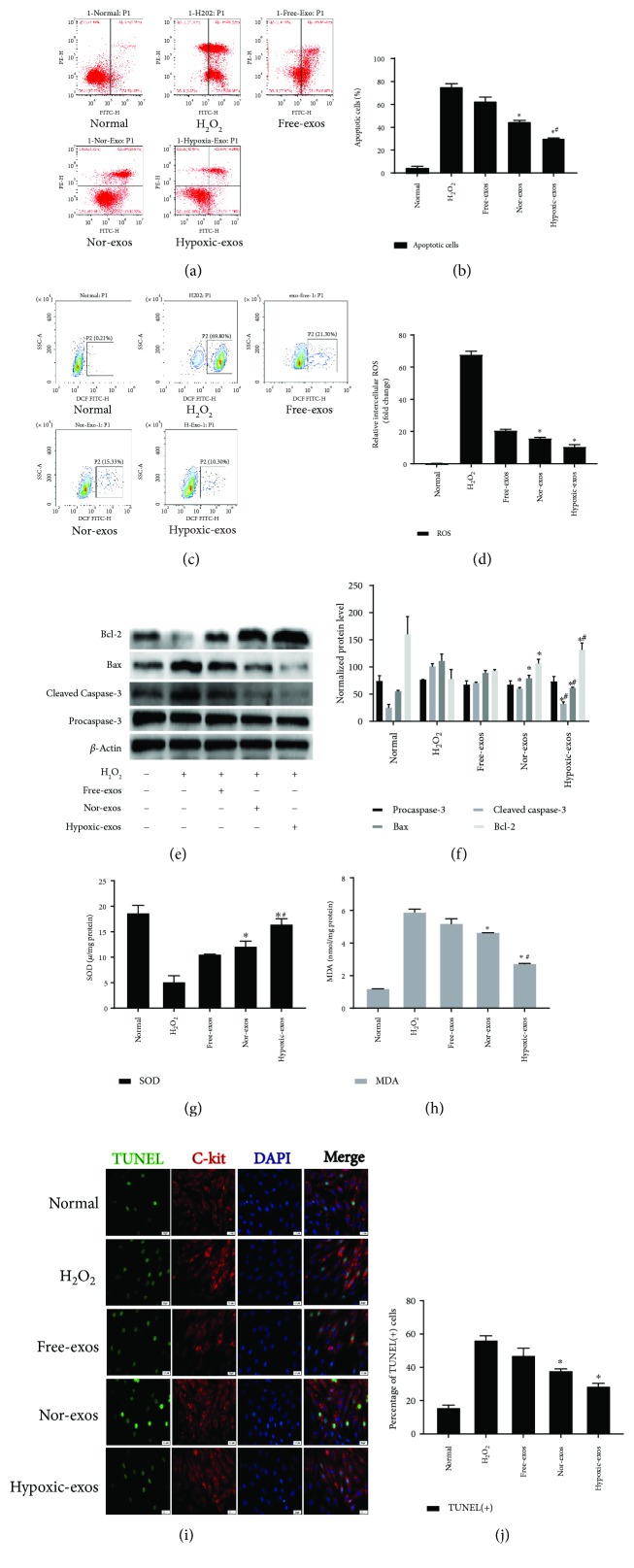
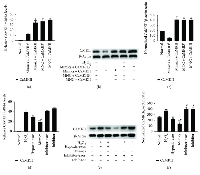
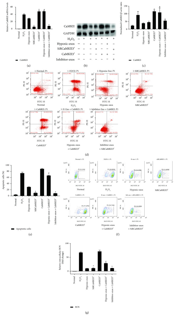
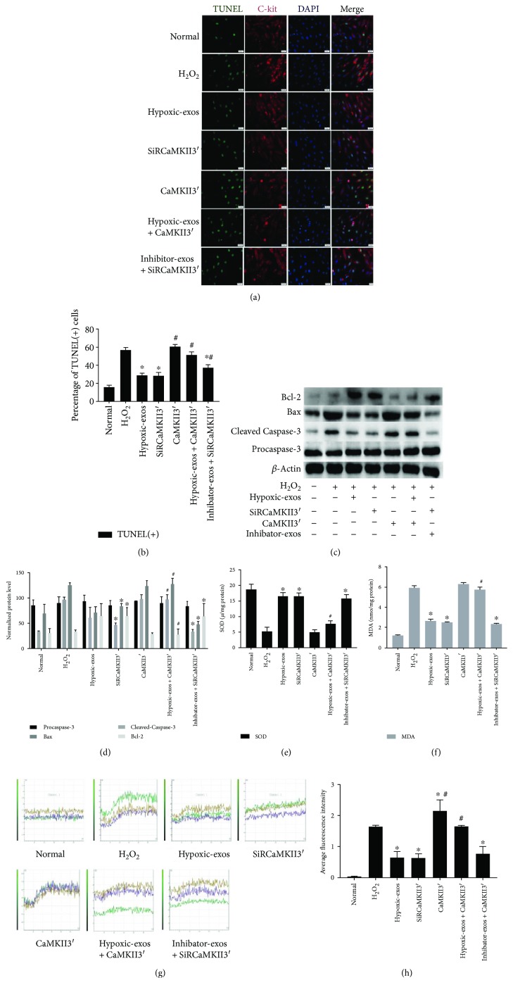
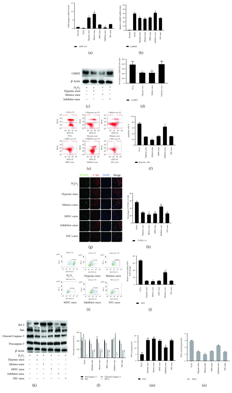
Cardiac stem cells (CSCs) have emerged as one of the most promising stem cells for cardiac protection. Recently, exosomes from bone marrow-derived mesenchymal stem cells (BMSCs) have been found to facilitate cell proliferation and survival by transporting various bioactive molecules, including microRNAs (miRs). In this study, we found that BMSC-derived exosomes (BMSC-exos) significantly decreased apoptosis rates and reactive oxygen species (ROS) production in CSCs after oxidative stress injury. Moreover, a stronger effect was induced by exosomes collected from BMSCs cultured under hypoxic conditions (Hypoxic-exos) than those collected from BMSCs cultured under normal conditions (Nor-exos). We also observed greater miR-214 enrichment in Hypoxic-exos than in Nor-exos. In addition, a miR-214 inhibitor or mimics added to modulate miR-214 levels in BMSC-exos revealed that exosomes from miR-214-depleted BMSCs partially reversed the effects of hypoxia-induced exosomes on oxidative damage in CSCs. These data further confirmed that miR-214 is the main effector molecule in BMSC-exos that protects CSCs from oxidative damage. miR-214 mimic and inhibitor transfection assays verified that CaMKII is a target gene of miR-214 in CSCs, with exosome-pretreated CSCs exhibiting increased miR-214 levels but decreased CaMKII levels. Therefore, the miR-214/CaMKII axis regulates oxidative stress-related injury in CSCs, such as apoptosis, calcium homeostasis disequilibrium, and excessive ROS accumulation. Collectively, these findings suggest that BMSCs release miR-214-containing exosomes to suppress oxidative stress injury in CSCs through CaMKII silencing.


Figures

Figure 1
Figure 1
Figure 2
Figure 2
Characterization of C-kit+ CSCs, exosomes, and cellular internalization. (a) Purified cells were double stained for c-kit (green) and DAPI (blue) and observed under a fluorescence microscope (Olympus, Japan). (b) Representative FCM characterization of C-kit+ CSCs for typical surface antigens and isotype control after magnetic bead sorting. Surface expression of C-kit, and the absence of surface expression of CD45 and CD34. (c) Transmission electron microscopy analysis of BMSC-exos. Scale bar = 100 nm. (d) NTA of exosome diameter and concentration. (e) Western blotting of the exosome markers CD63, CD9, and Alix. (f) CSCs were incubated with DiI-labeled BMSC-exos (400 μg/ml) for 24 h. Fluorescence photomicrographs showed internalized DiI-labeled BMSC-exos (red) in DAPI-labeled CSCs (blue). Scale bar = 20 μm.
Figure 3
Figure 3
Effect of miR-214 expression on CSC apoptosis under oxidative stress. Cells were treated with miR-214 mimics, inhibitors, or negative control RNA for 48 h and/or pretreated with BMSC-exos (400 μg/ml) for 24 h and then cultured with 100 μM H2O2 for 2 h for subsequent analyses. (a) RT-qPCR was used to analyze miR-214 expression in exosomes after normoxic or hypoxic preconditioning. Compared that in Nor-exos, miR-214 expression was significantly upregulated in Hypoxic-exos. (b) RT-qPCR analysis of miR-214 expression in CSCs after different treatments. Compared with that in the H2O2 group, miR-214 significantly upregulated in the Hypoxic-exos or miR-214 mimics group. Compared with the Hypoxic-exos group, the inhibitor-exos group displayed a significantly decreased miR-214 expression. (c) Representative dot plots of cell apoptosis after Annexin V/PI dual staining are shown. The upper left quadrant (% gated) shows necrotic cells (Annexin V−/PI+); the upper right quadrant (% fated) shows late apoptotic cells (Annexin V+/PI+); the left lower quadrant (% gated) shows live cells (Annexin V−/PI−); and the right lower quadrant (% gated) shows early apoptotic cells (Annexin V+/PI−). These cells were measured for comparison. (d) The percentage of apoptotic cells represents both early and late apoptotic cells. Compared with H2O2 or miR-214 inhibitor, Hypoxic-exos or miR-214 mimics decreased the percentage of apoptotic cells. In addition, the Hypoxic-exo-induced protective effect against CSC apoptosis under oxidative stress was partially suppressed by miR-214 inhibitors. (e) Representative immunofluorescence staining for TUNEL (green), C-kit (red), DAPI (blue), and merged images. Photos were randomly captured using a fluorescence microscope. Scale bar = 20 μm. (f) The panel shows the percentage of TUNEL-positive cells. Compared with H2O2 or miR-214 inhibitors, Hypoxic-exos or miR-214 mimics could significantly decrease the percentage of TUNEL-positive cells. In addition, compared with Hypoxic-exos, inhibitors-exos could partially increase the percentage of TUNEL-positive cells. (g) The intracellular ROS level was determined by FCM. The P2 percentage indicates the proportion of cells with increased ROS production, with signals above background DCF fluorescence levels. (h) Compared with that in CSCs treated with H2O2 or miR-214 inhibitors, the fluorescence intensity of intracellular ROS was decreased in CSCs treated with Hypoxic-exos or miR-214 mimics. Inhibitor-exos showed higher ROS fluorescence intensity than Hypoxic-exos. (i and j) Graph represents the SOD and MDA levels in CSCs, compared with H2O2 group, Hypoxic-exos or miR-214 mimics inhibited MDA levels and increased SOD production, while miR-214 inhibitors or inhibitor-exos increased MDA levels and suppressed SOD production. (k and l) The expression of apoptosis-related proteins, such as procaspase-3, cleaved caspase-3, Bax, and Bcl-2 were detected using immunoblotting. Compared with H2O2-treated cells, the cells treated with Hypoxic-exos or miR-214 mimics displayed substantially decreased cleaved caspase-3 and Bax expression and increased Bcl-2 expression. However, compared with Hypoxic-exos, miR-214 inhibitors or inhibitor-exos significantly increased cleaved caspase-3 and Bax expression but decreased Bcl-2 expression, n = 3; P < 0.05 compared with the H2O2 group; #P < 0.05 compared with the Hypoxic-exos group.
Figure 4
Figure 4
Exosomes released from hypoxia-pretreated BMSCs protect CSCs from oxidative stress injury. CSCs cultured with 100 μM H2O2 were pretreated with BMSC-exos (400 μg/ml) for 24 h and then subjected to analysis. (a) Representative dot plots of cell apoptosis after Annexin V/PI dual staining are shown. The left upper quadrant (% gated) shows necrotic cells (Annexin V−/PI+); the upper right quadrant (% gated) shows late apoptotic cells (Annexin V+/PI+); the left lower quadrant (% gated) shows live cells (Annexin V−/PI−); and the right lower quadrant (% gated) shows early apoptotic cells (Annexin V+/PI−). These cells were measured for comparison. (b) The percentage of apoptotic cells represents both early and late apoptotic cells. Compared with the H2O2-treated group, the BMSC-exo-treated group displayed a decreased percentage of apoptotic cells. In addition, Hypoxic-exos more markedly decreased apoptosis than did Nor-exos or Free-exos. (c) The intracellular ROS level was determined by FCM. The P2 percentage indicates the proportion of cells with increased ROS production, with signals above background 2′,7′-dichlorofluorescein (DCF) fluorescence levels. (d) Compared with the H2O2-treated group, the BMSC-exo-treated group had a significantly decreased intracellular ROS fluorescence intensity. In addition, Hypoxic-exos decreased ROS fluorescence to a greater degree than did Nor-exos or Free-exos. (e and f) The effects of BMSC-exos on cell apoptosis-related genes, such as procaspase-3, cleaved caspase-3, Bax, and Bcl-2 were detected by immunoblotting. Compared with the H2O2-treated cells, the BMSC-exo-treated cells had substantially decreased levels of cleaved caspase-3 and Bax and increased levels of Bcl-2. Additionally, Hypoxic-exos more markedly affected these protein levels than did Nor-exos. (g and h) Graph represents the SOD and MDA levels in CSCs; compared with H2O2 group, Hypoxic-exos inhibited MDA levels and increased SOD production. (i) Representative immunofluorescence staining for TUNEL (green), C-kit (red), DAPI (blue), and merged images. Photos were randomly captured using a fluorescence microscope. Scale bar = 20 μm. (j) The panel shows the percentage of TUNEL-positive cells. Compared with the H2O2-treated group, the BMSC-exo-treated group had significantly decreased percentage of TUNEL-positive cells. n = 3; P < 0.05 compared with the H2O2 group; #P < 0.05 compared with the Nor-exos group.
Figure 5
Figure 5
CaMKII is a target gene of miR-214 in CSCs. Cultured CSCs were transfected with CaMKII overexpression cDNA with or without the 3′UTR for 48 h. Subsequently, the cells were transfected with miR-214 mimics, inhibitors, or negative control RNA for 48 h. The cells were harvested and subjected to RT-qPCR or Western blotting analysis after treatment with BMSC-exos collected under different conditions for 24 h and/or cultured with 100 μM H2O2 for 2 h. (a) RT-qPCR analysis of CaMKII expression in CSCs after different treatments. After overexpressing cDNA for CaMKII containing the 3′UTR (CaMKII3′) in CSCs, CaMKII3′ mRNA levels dramatically decreased in response to treatment with miR-21 mimics as demonstrated by RT-qPCR. However, miR-214 mimics had no effect on mRNA levels of CaMKII without the 3′UTR. (b-c) CaMKII protein levels were detected by immunoblotting. miR-214 mimics could significantly downregulate the expression of CaMKII with the 3′UTR at protein levels. However, miR-214 mimics had no effect on the protein levels of CaMKII without the 3′UTR. n = 3; P < 0.05 compared with the mimics + CaMKII3′ group. (d) RT-qPCR analysis of CaMKII expression in CSCs after different treatments. Compared with that in the normal group, the CaMKII mRNA level was significantly upregulated in the H2O2 group. Compared with H2O2, Hypoxic-exos or miR-214 mimics significantly suppressed CaMKII mRNA expression. (e-f) Western blotting was used to verify the effect of exosomal miR-214 on CaMKII expression in CSCs. CaMKII protein levels were dramatically decreased after Hypoxic-exo or mimic-exo (exosomes from miR-214-mimic-modified BMSCs) treatment relative to those with H2O2 treatment. However, compared with Hypoxic-exos, miR-214 inhibitors or inhibitor-exos upregulated CaMKII protein levels. n = 3; P < 0.05 compared with the H2O2 group; #P < 0.05 compared with the Hypoxic-exos group.
Figure 6
Figure 6
Change in CaMKII expression during BMSC-exo-induced antiapoptotic effect in CSCs under oxidative stress. Cultured CSCs were transfected with CaMKII3′ overexpression cDNA or siRCaMKII3′ for 48 h. Then, the cells were treated with BMSC-exos under different conditions for 24 h and/or cultured with 100 μM H2O2 for 2 h. (a) RT-qPCR were carried out to detect CaMKII mRNA levels. Compared with other treatments, CaMKII3′ transfection significantly upregulated CaMKII expression, and SiRCaMKII3′ transfection significantly downregulated CaMKII mRNA levels in CSCs. (b-c) Western blotting revealed that compared with H2O2 treatment, CaMKII3′ transfection upregulated CaMKII protein levels, while SiRCaMKII3′ transfection downregulated CaMKII protein levels in CSCs. Additionally, compared with Hypoxic-exos, Hypoxic-exos + CaMKII3′ upregulated CaMKII protein levels. (d) Representative dot plots of cell apoptosis after Annexin V/PI dual staining are shown. The upper left quadrant (% gated) shows necrotic cells (Annexin V−/PI+); the upper right quadrant (% gated) shows late apoptotic cells (Annexin V+/PI+); the left lower quadrant (% gated) shows live cells (Annexin V−/PI−); and the right lower quadrant (% gated) shows early apoptotic cells (Annexin V+/PI-). These cells were measured for comparison. (e) The percentage of apoptotic cells represents both early and late apoptotic cells. Compared with the H2O2 group, the SiRCaMKII3′-transfected group displayed a decreased percentage of apoptotic cells. In addition, the Hypoxic-exo-induced protective effect on CSC apoptosis under oxidative stress was suppressed by CaMKII3′ overexpression. (f) Intracellular ROS level was determined by FCM. The P2 percentage indicates the proportion of cells with increased ROS production, with fluorescence levels above background DCF fluorescence levels. (g) Compared with that in H2O2-treated CSCs, fluorescence intensity of intracellular ROS was decreased in SiRCaMKII3′-treated CSCs. In addition, the Hypoxic-exo-induced protective effect on CSCs against oxidative stress injury was suppressed by CaMKII3′ overexpression. n = 3; P < 0.05 compared with the H2O2 group. #P < 0.05 compared with Hypoxic-exos group.
Figure 7
Figure 7
Change in CaMKII expression during BMSC-exo-induced antioxidative injury in CSCs under oxidative stress. Cultured CSCs were transfected CaMKII3′ overexpression cDNA or siRCaMKII3′ for 48 h. Then, the cells were treated with BMSC-exos under different conditions for 24 h and/or cultured with 100 μM H2O2 for 2 h. (a) Representative immunofluorescence staining for TUNEL (green), C-kit (red), DAPI (blue), and merged images. Photos were randomly captured using a fluorescence microscope. Scale bar = 20 μm. (b) The panel shows the percentages of TUNEL-positive cells. Compared with H2O2, SiRCaMKII3′ could significantly decreased the percentage of TUNEL-positive cells. Additionally, compared with Hypoxic-exos, CaMKII3′ could partially increase the percentage of TUNEL-positive cells. (c and d) The expression levels of procaspase-3, cleaved caspase-3, Bax, and Bcl-2 were detected by immunoblotting. Compared with Hypoxic-exos or SiRCaMKII3′ group, the CaMKII3′ group displayed substantially increased cleaved caspase-3 and Bax expression and decreased Bcl-2 expression. In addition, the Hypoxic-exo-induced protective effect against CSC apoptosis under oxidative stress was suppressed by CaMKII3′ overexpression. (e and f) Graph represents the SOD and MDA levels in CSCs; compared with H2O2 group, Hypoxic-exos or SiRCaMKII3′ inhibited MDA levels and increased SOD production, while CSCs were transfected with CaMKII3′ increased MDA levels and suppressed SOD production. (g) Transient intracellular Ca2+ measurement assays with Fluo-8/AM fluorescent labeling were used to detect Ca2+ concentration in CSCs exposed to different treatments. (h) Compared with that in the H2O2 or CaMKII3′ group, the fluorescence intensity of intracellular Ca2+ was significantly decreased in the Hypoxic-exos or siRCaMKII group. Furthermore, CaMKII3′ overexpression could suppress the Hypoxic-exo-induced protective effect against CSC oxidative stress injury. n = 3; P < 0.05 compared with the H2O2 group. #P < 0.05 compared with the Hypoxic-exos group.
Figure 8
Figure 8
Exosomes derived from miR-214-modified BMSCs exert an antiapoptotic effect on CSCs under oxidative stress. BMSCs were transfected with miR-214 mimics, inhibitors, or negative control RNA. At 48 h posttransfection, exosomes were isolated from BMSCs pretreated with hypoxia and then added to CSCs under oxidative stress for 2 h. (a) RT-qPCR analysis of miR-214 expression in CSCs after different treatments. Compared with that in the H2O2 group, miR-214 was significantly upregulated in mimic-exos group and substantially downregulated in the inhibitor-exos group. (b) RT-qPCR analysis of CaMKII mRNA expression in CSCs after different treatments. Compared with that in the H2O2 group, CaMKII mRNA was significantly downregulated in the mimic-exos group but substantially upregulated in the inhibitor-exos group. (c and d) Western blotting was carried out to detect CaMKII protein levels, which revealed that compared with those in H2O2-treated CSCs, CaMKII levels were significantly downregulated in Hypoxic-exo- or mimic-exo-treated CSCs, whereas CaMKII protein levels were significantly upregulated in inhibitor-exo-treated CSCs. (e) Representative cell apoptosis dot plots after Annexin V/PI dual staining are shown. The upper left quadrant (% gated) shows necrotic cells (Annexin V−/PI+); the upper right quadrant (% gated) shows late apoptotic cells (Annexin V+/PI+); the left lower quadrant (% gated) shows live cells (Annexin V−/PI−); and the right lower quadrant (% gated) shows early apoptotic cells (Annexin V+/PI−). These cells were measured for comparison. (f) The percentage of apoptotic cells represents both early and late apoptotic cells. Compared with the H2O2 group, the Hypoxic-exos or mimic-exos group displayed a decreased percentage of apoptotic cells. In addition, compared with Hypoxic-exos, inhibitor-exos increased the percentage of apoptotic cells. (g) Representative immunofluorescence staining for TUNEL (green), C-kit (red), DAPI (blue), and merged images. Photos were randomly captured using a fluorescence microscope. Scale bar = 20 μm. (h) The panel shows the percentage of TUNEL-positive cells. Compared with H2O2, Hypoxic-exos or mimic-exos could significantly decrease the percentage of TUNEL-positive cells. Additionally, compared with Hypoxic-exos, inhibitor-exos could partially increase the percentage of TUNEL-positive cells. (i) The intracellular ROS level was determined by FCM. The P2 percentage indicates the proportion of cells with increased ROS production, with signals above background DCF fluorescence levels. (j) Compared with that in H2O2-treated CSCs, the fluorescence intensity of intracellular ROS was decreased in Hypoxic-exo- or mimic-exo-treated CSCs. However, compared with Hypoxic-exos, inhibitor-exos decreased the fluorescence intensity of intracellular ROS in CSCs. (k and l) The expression levels of procaspase-3, cleaved caspase-3, Bax, and Bcl-2 were detected by immunoblotting. Compared with H2O2 or inhibitor-exos, Hypoxic-exos or mimic-exos substantially decreased cleaved caspase-3 and Bax expression and increased Bcl-2 expression. (m and n) Graph represents the SOD and MDA levels in CSCs; compared with H2O2 group, Hypoxic-exos or mimics-exos inhibited MDA levels and increased SOD production, while inhibitor-exos group increased MDA levels and suppressed SOD production. n = 3; P < 0.05 compared with the H2O2 group; #P < 0.05 compared with the Hypoxic-exos group.

Similar articles

Cited by

References

    1. Beltrami A. P., Barlucchi L., Torella D., et al. Adult cardiac stem cells are multipotent and support myocardial regeneration. Cell. 2003;114(6):763–776. doi: 10.1016/S0092-8674(03)00687-1. - DOI - PubMed
    1. Mayfield A. E., Tilokee E. L., Davis D. R. Resident cardiac stem cells and their role in stem cell therapies for myocardial repair. The Canadian Journal of Cardiology. 2014;30(11):1288–1298. doi: 10.1016/j.cjca.2014.03.018. - DOI - PubMed
    1. Hong K. U., Bolli R. Cardiac stem cell therapy for cardiac repair. Current Treatment Options in Cardiovascular Medicine. 2014;16(7):p. 324. doi: 10.1007/s11936-014-0324-3. - DOI - PMC - PubMed
    1. Bolli R., Tang X. L., Sanganalmath S. K., et al. Intracoronary delivery of autologous cardiac stem cells improves cardiac function in a porcine model of chronic ischemic cardiomyopathy. Circulation. 2013;128(2):122–131. doi: 10.1161/CIRCULATIONAHA.112.001075. - DOI - PMC - PubMed
    1. Tang X. L., Li Q., Rokosh G., et al. Long-term outcome of administration of c-kitPOS cardiac progenitor cells after acute myocardial infarction: transplanted cells do not become cardiomyocytes, but structural and functional improvement and proliferation of endogenous cells persist for at least one year. Circulation Research. 2016;118(7):1091–1105. doi: 10.1161/CIRCRESAHA.115.307647. - DOI - PMC - PubMed

MeSH terms

Substances

LinkOut - more resources