

Effectiveness of modified dural incision to preserve the patency of the occipital sinus in foramen magnum decompression for a patient with Chiari malformation type I
- PMID: 30159197
- PMCID: PMC6094497
- DOI: 10.4103/sni.sni_70_18
Effectiveness of modified dural incision to preserve the patency of the occipital sinus in foramen magnum decompression for a patient with Chiari malformation type I
Abstract
Background: Foramen magnum decompression (FMD) has been acknowledged as a standard surgical procedure for symptomatic patients with Chiari malformation type I (CM-I). However, even if dural incision is necessary during FMD, the procedure of cutting off the occipital sinus has not been regarded as a safe option.
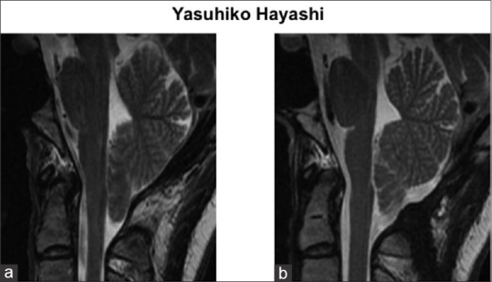
Case description: A 27-year-old woman with intractable occipital headache was diagnosed with CM-I without syringomyelia. Preoperative examination revealed a large oblique occipital sinus on her right side. During the first FMD, the dura mater was not incised to preserve the occipital sinus. However, her headache was not relieved with painkillers and cerebellar tonsillar ectopia remained. During the second FMD, two dural incisions were made, while preserving the occipital sinus patency. The dural patch was made using an autologous fascia for both dural incisions. Postoperatively, headache was completely resolved immediately, and cerebellar tonsil was elevated without any complication.
Conclusion: This dural incision, which is a modification of the method introduced by Pritz, would be a useful FMD option for patients of CM-I with dominant occipital sinus, which would lead to the serious neurological sequelae if the sinus flow is disturbed.
Keywords: Chiari malformation; dura; foramen magnum decompression; incision; occipital sinus.
Conflict of interest statement
There are no conflicts of interest.
Figures


References
-
- Alperin N, Loftus JR, Bagci AM, Lee SH, Oliu CJ, Shah AH, Green BA. Magnetic resonance imaging-based measures predictive of short-term surgical outcome in patients with Chiari malformation Type I: A pilot study. J Neurosurg Spine. 2017;26:28–38. - PubMed
-
- Arnautovic A, Splavski B, Boop FA, Arnautovic KI. Pediatric and adult Chiari malformation Type I surgical series 1955-2013: A review of demographics, operative treatment, and outcomes. J Neurosurg Pediatr. 2015;15:161–77. - PubMed
-
- Beyrouti R, Mansour M, Kacem A, Zaouali J, Mrissa R. Occipital sinus thrombosis: An exceptional case report. J Stroke Cerebrovasc Dis. 2016;25:e71–3. - PubMed
-
- Das AC, Hassan M. The occipital sinus. J Neurosurg. 1970;33:307–11. - PubMed
LinkOut - more resources
Full Text Sources
Other Literature Sources