

Conduction Aphasia as Initial Manifestation of Tuberculous Meningitis
- PMID: 30159215
- PMCID: PMC6110626
- DOI: 10.7759/cureus.2889
Conduction Aphasia as Initial Manifestation of Tuberculous Meningitis
Abstract
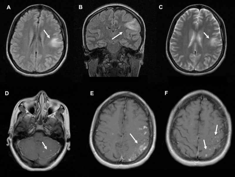
Conduction aphasia being the arcuate fasciculus of the site of structural injury is a speech disorder characterized by fluent, spontaneous speech and paraphasias, intact auditory comprehension, and limited repetition. One of the causes of stroke in young adults is the Mycobacterium tuberculosis (MTB) infection, which may cause cerebral ischemia secondary to artery obliteration. In this case report, we present a previously healthy 24-year-old woman that presented with a sudden onset of aphasia; MTB was identified as the etiological agent. Tuberculous meningitis (TBM) has a wide range of clinical manifestations with aphasia being one of the rarest forms of initial presentation.
Keywords: aphasia; conduction aphasia; mycobacterium tuberculosis; stroke; tuberculous meningitis.
Conflict of interest statement
The authors have declared that no competing interests exist.
Figures
Similar articles
-
The Role of Early Rehabilitation in Better Outcomes in a Rare Presentation of Tuberculous Meningitis With Broca's Aphasia.Cureus. 2024 Feb 7;16(2):e53793. doi: 10.7759/cureus.53793. eCollection 2024 Feb. Cureus. 2024. PMID: 38465188 Free PMC article.
-
Conduction aphasia in a 3-year-old with a left posterior cortical/subcortical abscess.Brain Lang. 1998 Mar;62(1):70-88. doi: 10.1006/brln.1998.1888. Brain Lang. 1998. PMID: 9570880
-
Diffusion tensor imaging depicting damage to the arcuate fasciculus in patients with conduction aphasia: a study of the Wernicke-Geschwind model.Neurol Res. 2010 Sep;32(7):775-8. doi: 10.1179/016164109X12478302362653. Epub 2009 Oct 12. Neurol Res. 2010. PMID: 19825277
-
Post-stroke language disorders.Acta Clin Croat. 2011 Mar;50(1):79-94. Acta Clin Croat. 2011. PMID: 22034787 Review.
-
The Efficacy and Safety of Pharmacological Treatments for Post-stroke Aphasia.CNS Neurol Disord Drug Targets. 2018;17(7):509-521. doi: 10.2174/1871527317666180706143051. CNS Neurol Disord Drug Targets. 2018. PMID: 29984673
Cited by
-
The Role of Early Rehabilitation in Better Outcomes in a Rare Presentation of Tuberculous Meningitis With Broca's Aphasia.Cureus. 2024 Feb 7;16(2):e53793. doi: 10.7759/cureus.53793. eCollection 2024 Feb. Cureus. 2024. PMID: 38465188 Free PMC article.
References
Publication types
LinkOut - more resources
Full Text Sources
Other Literature Sources