Dural Arteriovenous Fistula Associated With a Vestibular Tumor: An Unusual Case and Review of the Literature
- PMID: 30159216
- PMCID: PMC6110627
- DOI: 10.7759/cureus.2890
Dural Arteriovenous Fistula Associated With a Vestibular Tumor: An Unusual Case and Review of the Literature
Abstract
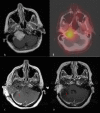
Intracranial dural arteriovenous fistulae (DAVF) are rare vascular malformations. They are generally considered to be acquired lesions, often attributed to dural sinus thrombosis and intracranial venous hypertension. The authors encountered a case of DAVF associated with an octreotide-positive vestibular schwannoma. A 46-year-old female had symptoms of right ear congestion accompanied by pulsatile tinnitus and mild hearing loss. Magnetic resonance imaging (MRI) identified a lobulated mass centered at the cerebellopontine angle. Preoperatively, on cerebral angiography, there was an incidental discovery of a DAVF in the right posterior fossa. The decision was made to proceed with resection of the tumor in a staged fashion. Her latest follow-up MRI showed no evidence of recurrent tumor. This is the second reported case of DAVF associated with an intracranial schwannoma. Findings are discussed along with a thorough review of the literature. This case, combined with the data from the literature review, led us to believe that tumor-related angiogenesis might contribute to DAVF formation.
Keywords: angiogenesis; dural arteriovenous fistula; intracranial tumor; octreotide scintigraphy; somatostatin receptor; vestibular schwannoma.
Conflict of interest statement
The authors have declared that no competing interests exist.
Figures


Similar articles
-
Ruptured dural arteriovenous fistula and sinus venous thrombosis following surgical resection of a vestibular schwannoma: Case report and review of the literature.Neurochirurgie. 2022 Dec;68(6):688-692. doi: 10.1016/j.neuchi.2022.03.008. Epub 2022 May 20. Neurochirurgie. 2022. PMID: 35599062 Review.
-
Optic neuropathy, a warning for cerebral venous sinus thrombosis and underlying dural arteriovenous fistulae.J Int Med Res. 2022 Jul;50(7):3000605221078071. doi: 10.1177/03000605221078071. J Int Med Res. 2022. PMID: 35899780 Free PMC article.
-
Development of a dural arteriovenous fistula subsequent to cerebral venous thrombosis by venous hypertension.eNeurologicalSci. 2018 Nov 22;14:24-27. doi: 10.1016/j.ensci.2018.11.015. eCollection 2019 Mar. eNeurologicalSci. 2018. PMID: 30555948 Free PMC article.
-
Dural arteriovenous fistula associated with intratumor hemorrhage.J Clin Neurosci. 2019 Jan;59:352-355. doi: 10.1016/j.jocn.2018.10.008. Epub 2018 Oct 31. J Clin Neurosci. 2019. PMID: 30391309
-
[Multiple dural arteriovenous fistulas involving both the cavernous sinus and the posterior fossa: report of two cases and review of the literature].No Shinkei Geka. 2001 Nov;29(11):1065-72. No Shinkei Geka. 2001. PMID: 11758314 Review. Japanese.
Cited by
-
Low-Frequency Air-Bone Gap and Pulsatile Tinnitus Due to a Dural Arteriovenous Fistula: Considerations upon Possible Pathomechanisms and Literature Review.Audiol Res. 2023 Nov 1;13(6):833-844. doi: 10.3390/audiolres13060073. Audiol Res. 2023. PMID: 37987331 Free PMC article.
-
The Spinal Dural Arteriovenous Fistula in a Patient With Metastatic Renal Cell Carcinoma.Cureus. 2021 May 28;13(5):e15303. doi: 10.7759/cureus.15303. Cureus. 2021. PMID: 34221759 Free PMC article.
References
-
- Cerebral dural arteriovenous fistulas: clinical and angiographic correlation with a revised classification of venous drainage. Cognard C, Gobin YP, Pierot L, et al. Radiology. 1995;194:671–680. - PubMed
-
- Dural arteriovenous malformations of the transverse/sigmoid sinus acquired from dominant sinus occlusion by a tumor: report of two cases. Arnautovic KI, Al-Mefty O, Angtuaco E, Phares LJ. Neurosurgery. 1998;42:383–388. - PubMed
-
- Intracranial dural arteriovenous fistula associated with a spinal tumor: a case report. Hatanaka R, Takai K, Iijima A, Taniguchi M. Acta Neurochir (Wien) 2015;157:1825–1827. - PubMed
-
- Dural arteriovenous fistula associated with meningioma: spontaneous disappearance after tumor removal. Ahn JY, Lee BH, Cho YJ, Joo JY, Lee KS. Neurol Med Chir (Tokyo) 2003;43:308–311. - PubMed
Publication types
LinkOut - more resources
Full Text Sources
Other Literature Sources
