

Erythema Sweetobullosum: A Reactive Cutaneous Manifestation of Coccidioidomycosis
- PMID: 30159356
- PMCID: PMC6109837
- DOI: 10.1177/2324709618796659
Erythema Sweetobullosum: A Reactive Cutaneous Manifestation of Coccidioidomycosis
Abstract
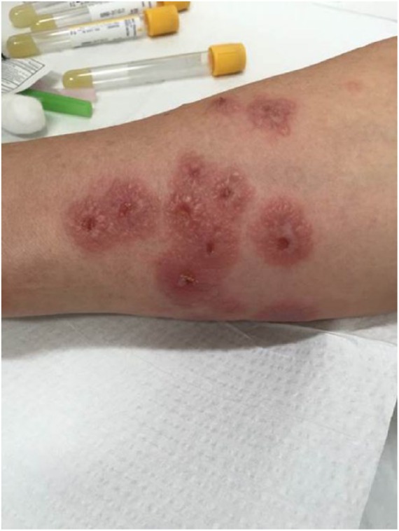
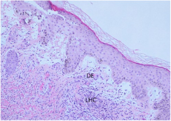
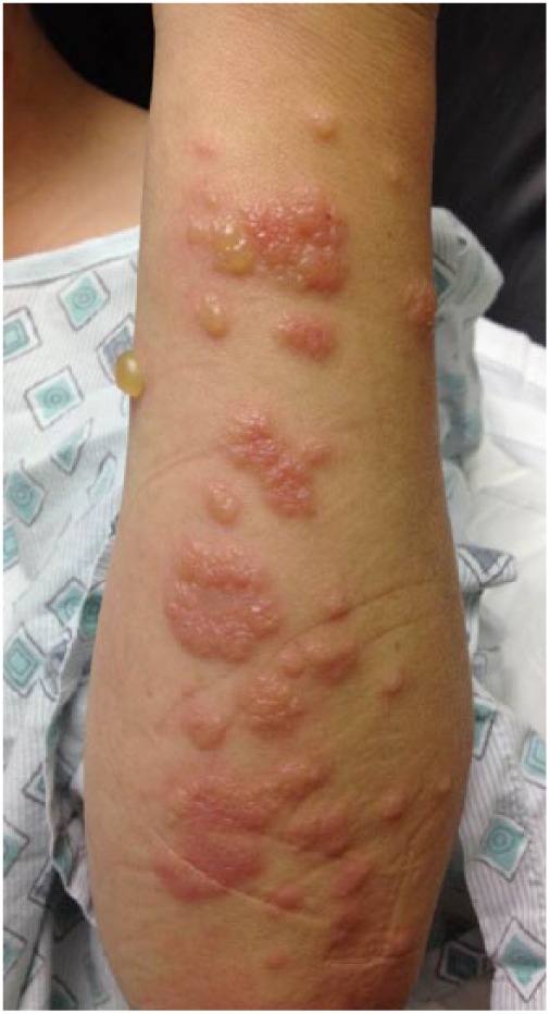
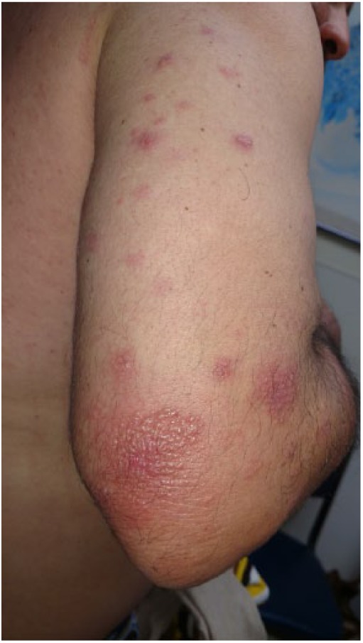
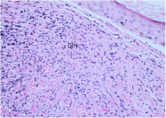
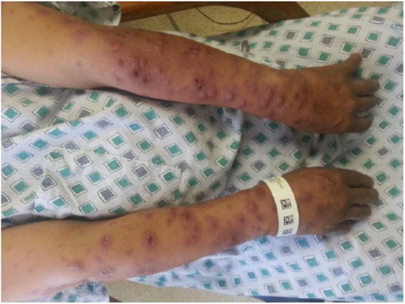
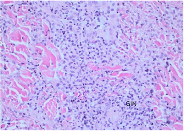
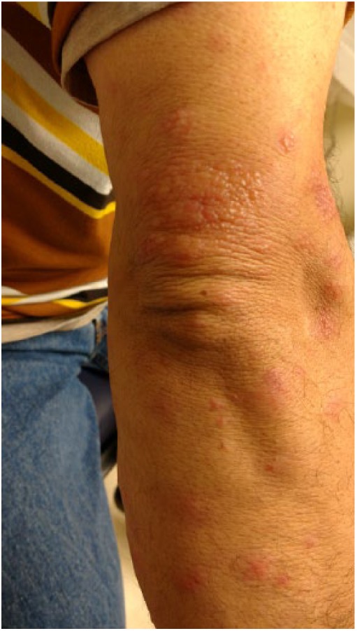
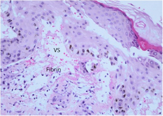
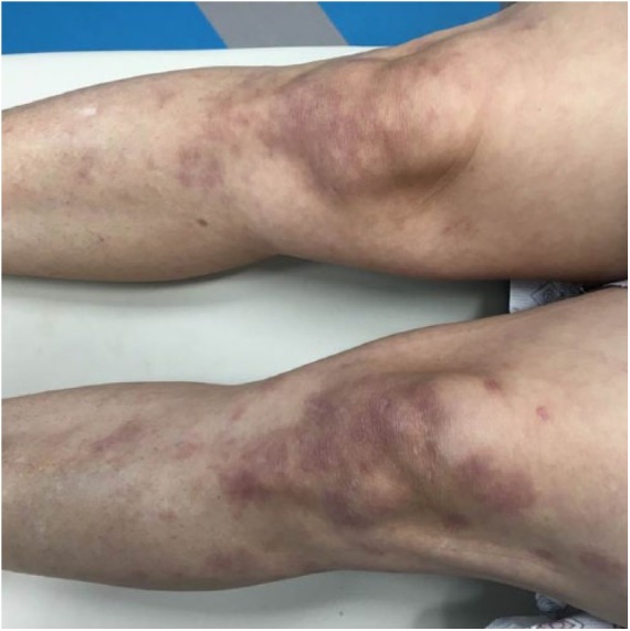
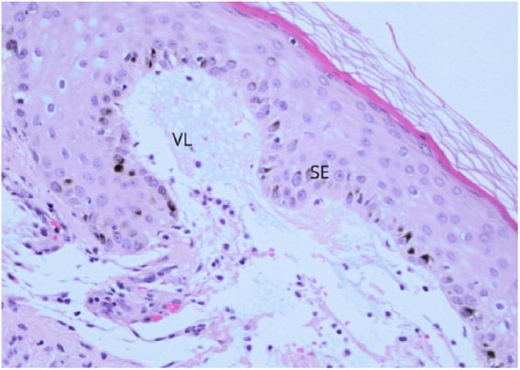
Reactive cutaneous coccidioidal skin manifestations are commonly noticed during the early stage of coccidioidomycosis. These skin lesions are devoid of any active coccidioidal organism, and the immune trigger mechanisms are not elucidated. We describe 6 cases of unusual reactive cutaneous coccidioidal manifestation, characterized by painful vesiculobullous lesions known as erythema sweetobullosum. The biopsy of the lesions revealed neutrophilic dermatosis with inflammatory cells resulting in a cleft and elevation of the most superficial layer of the skin forming a bulla. The reactive cutaneous lesion is self-limited and requires no specific therapy.
Keywords: coccidioidomycosis; cutaneous manifestation; erythema sweetobullosum; noninfectious.
Conflict of interest statement
Declaration of Conflicting Interests: The author(s) declared no potential conflicts of interest with respect to the research, authorship, and/or publication of this article.
Figures
Similar articles
-
State-of-the-art treatment of coccidioidomycosis: skin and soft-tissue infections.Ann N Y Acad Sci. 2007 Sep;1111:411-21. doi: 10.1196/annals.1406.010. Epub 2007 Mar 1. Ann N Y Acad Sci. 2007. PMID: 17332079 Review.
-
Diagnosis of disseminated coccidioidomycosis by liver biopsy.Arch Intern Med. 1983 Jul;143(7):1335-8. Arch Intern Med. 1983. PMID: 6870405
-
Erythema nodosum.Am Fam Physician. 1984 Oct;30(4):227-32. Am Fam Physician. 1984. PMID: 6496266
-
Neutrophilic dermatoses: a review of current treatment options.Am J Clin Dermatol. 2009;10(5):301-12. doi: 10.2165/11310730-000000000-00000. Am J Clin Dermatol. 2009. PMID: 19658442 Review.
-
Purpuric macules with vesiculobullous lesions: a novel manifestation of Chikungunya.Int J Dermatol. 2011 Jan;50(1):61-9. doi: 10.1111/j.1365-4632.2010.04644.x. Int J Dermatol. 2011. PMID: 21182504 Clinical Trial.
References
-
- Hospenthal DR, Rinaldi MG. Diagnosis and Treatment of Human Mycoses. Berlin, Germany: Springer Science + Business Media; 2007.
-
- Stevens DA. Coccidioidomycosis. N Engl J Med. 1995;332:1077-1082. - PubMed
LinkOut - more resources
Full Text Sources
Other Literature Sources
