

Longitudinal bleb morphology in anterior segment OCT after minimally invasive transscleral ab interno Glaucoma Gel Microstent implantation
- PMID: 30160048
- PMCID: PMC6586011
- DOI: 10.1111/aos.13902
Longitudinal bleb morphology in anterior segment OCT after minimally invasive transscleral ab interno Glaucoma Gel Microstent implantation
Abstract
Purpose: Like the classic trabeculectomy, the minimally invasive, ab interno XEN Glaucoma Gel Microstent (XEN-GGM) creates a filtration bleb in the conjunctiva. The goal of this study was to investigate internal bleb morphology over time with anterior segment optical coherence tomography (AS-OCT) after XEN-GGM implantation.
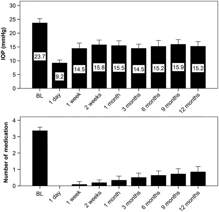
Methods: In a prospective, single-centre, single-armed cohort study, blebs were characterized using AS-OCT in 78 eyes of 60 patients at day 1, at weeks 1 and 2 and at months 1, 3, 6, 9 and 12 after XEN-GGM implantation in patients with open-angle glaucoma. Morphological bleb characteristics were correlated with IOP and surgical success.
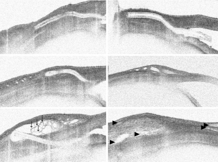
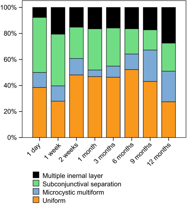
Results: Anterior segment optical coherence tomography data indicate early and late bleb changes in the course of 12 months. Uniform blebs in AS-OCTs showed higher IOPs at all examinations between week 1 (17.7 ± 4.8 mmHg versus 11.3 ± 7.1 mmHg, p = 0.001) and month 3 (16.4 ± 6.1 versus 13.4 ± 6.1, p = 0.04). Subconjunctival tissue separation bleb morphology was associated with lower mean IOPs during the course of 12 months (r = -0.75, p = 0.031). Predictors for surgical failure at month 12 were microcystic multiform bleb morphology in AS-OCT at month 3 (60% versus 15%, relative risk 4.0, p = 0.043) and uniform bleb morphology at month 9 (33% versus 23%, relative risk 1.4, p = 0.015).
Conclusion: Bleb appearance after XEN surgery seems to be different to classic trabeculectomy literature. The present data suggest correlation of IOP and surgical long-term success with bleb morphology in AS-OCT. Prevalence of small diffuse cysts is directly associated with lower IOPs, while cystic encapsulation at 3 months predicts higher surgical failure.
Keywords: XEN; glaucoma; minimally invasive glaucoma surgery; subconjunctival implant; surgical success.
© 2018 The Authors. Acta Ophthalmologica published by John Wiley & Sons Ltd on behalf of Acta Ophthalmologica Scandinavica Foundation.
Figures
References
-
- Azuara‐Blanco A & Katz LJ (1998): Dysfunctional filtering blebs. Surv Ophthalmol 43: 93–126. - PubMed
-
- Bouhenni RA, Al Jadaan I, Rassavong H, Al Shahwan S, Al Katan H, Dunmire J, Krasniqi M & Edward DP (2016): Lymphatic and blood vessel density in human conjunctiva after glaucoma filtration surgery. J Glaucoma 25: e35–e38. - PubMed
-
- Cairns JE (1968): Trabeculectomy. Preliminary report of a new method. Am J Ophthalmol 66: 673–679. - PubMed
-
- Cantor LB, Mantravadi A, WuDunn D, Swamynathan K & Cortes A (2003): Morphologic classification of filtering blebs after glaucoma filtration surgery: the Indiana Bleb Appearance Grading Scale. J Glaucoma 12: 266–271. - PubMed
-
- Conlon R, Saheb H & Ahmed II (2017): Glaucoma treatment trends: a review. Canadian Journal of Ophthalmology. Journal Canadien D'ophtalmologie 52: 114–124. - PubMed
Publication types
MeSH terms
Substances
Grants and funding
LinkOut - more resources
Full Text Sources
Other Literature Sources
Research Materials
