Review
. 2018 Aug 30;14(8):e1007479.
doi: 10.1371/journal.pgen.1007479. eCollection 2018 Aug.

PRDM9, a driver of the genetic map

Affiliations
Review

PRDM9, a driver of the genetic map

Corinne Grey et al. PLoS Genet. .

Abstract

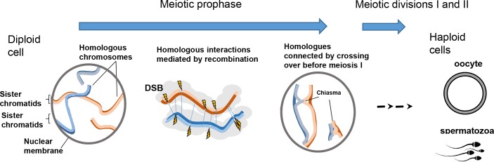
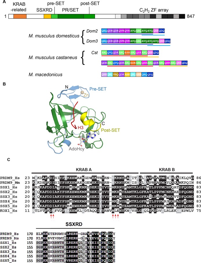
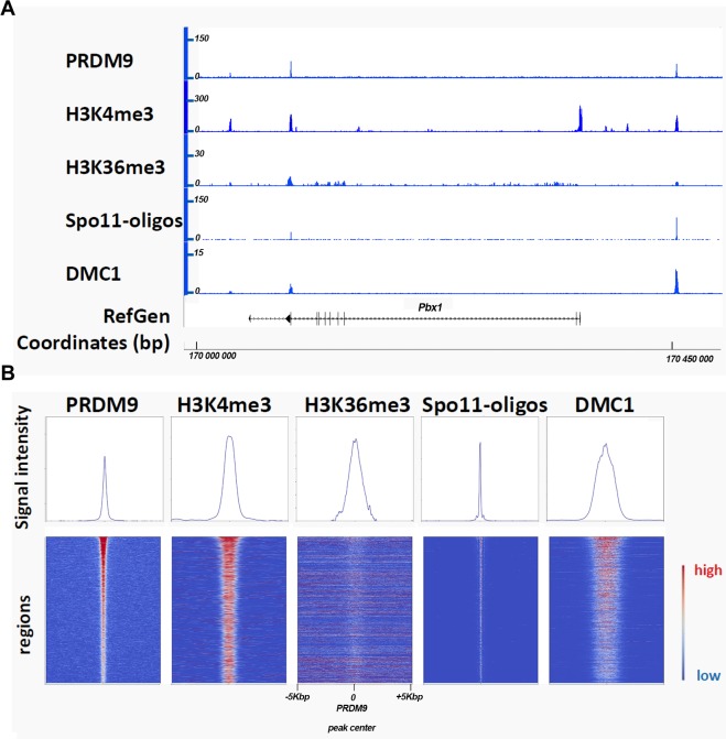
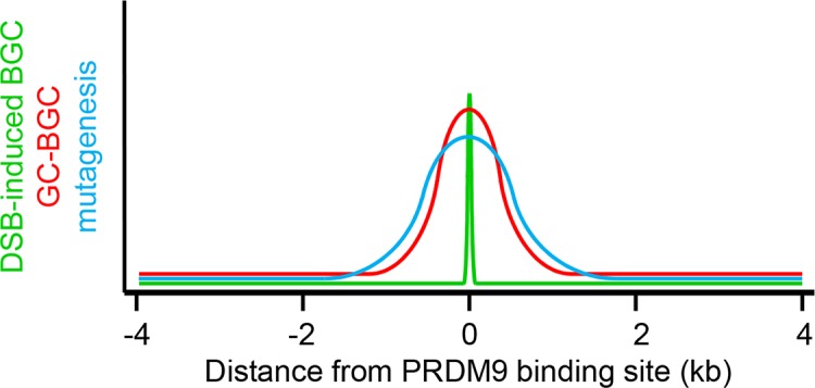
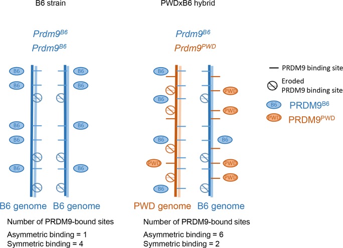
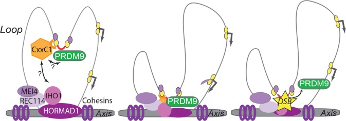
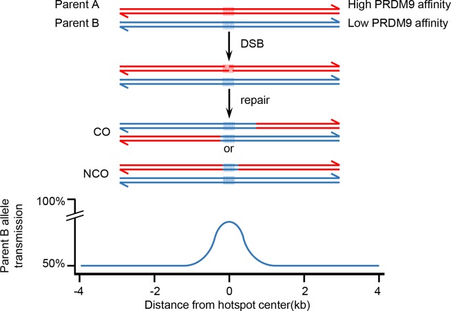
During meiosis, maternal and paternal chromosomes undergo exchanges by homologous recombination. This is essential for fertility and contributes to genome evolution. In many eukaryotes, sites of meiotic recombination, also called hotspots, are regions of accessible chromatin, but in many vertebrates, their location follows a distinct pattern and is specified by PR domain-containing protein 9 (PRDM9). The specification of meiotic recombination hotspots is achieved by the different activities of PRDM9: DNA binding, histone methyltransferase, and interaction with other proteins. Remarkably, PRDM9 activity leads to the erosion of its own binding sites and the rapid evolution of its DNA-binding domain. PRDM9 may also contribute to reproductive isolation, as it is involved in hybrid sterility potentially due to a reduction of its activity in specific heterozygous contexts.


Conflict of interest statement

The authors have declared that no competing interests exist.

Figures

Fig 1
Fig 1. The role of homologous recombination during meiotic prophase.
During meiotic prophase, homologous recombination allows the interaction, alignment, and connection through chiasmata of homologous chromosomes. Chromosomes are structured by a protein axis (blue and red lines for paternal and maternal chromosomes, respectively) to which chromatin loops (grey cloud) are anchored. The recombination pathway is initiated by the formation of several DSBs (yellow lightning) on each homologue. DSB sites are determined by PRDM9. DSB repair promotes interactions between homologues (thin blue dotted lines). A small subset of DSB repair events, at least one per homologue pair, leads to a crossover visualized as a chiasma that establishes a topological connection between homologues. These connections are required for proper chromosome segregation at the first meiotic division (meiosis I). The two meiotic divisions lead to the formation of haploid oocytes and spermatozoa. DSB, DNA double strand break; PRDM9, PR domain-containing protein 9.
Fig 2
Fig 2. PRDM9 domains.
(A) The domain organization of PRDM9 and the high diversity of its zinc finger array. Top, schematic representation of the mouse PRDM9 protein. Bottom, zinc finger arrays from two Mus musculus domesticus alleles (Dom2 and Dom3 from C57BL/6 and C3H lab strains, respectively), two M. m. castaneus alleles (including Cst from CAST strain) and one representative allele from Mus macedonicus (all alleles described in [31]) are shown underneath. Each box represents one zinc finger, and each color represents a specific zinc finger sequence. The DNA-contacting amino acids in position −1, 3, and 6 of the zinc finger are indicated within each box. Colored bars underline blocks of zinc fingers conserved between alleles from the same subspecies. The recurrence of several individual zinc fingers is apparent within each array. Alleles from the same species (M. musculus) are largely made of combinations of the same set of zinc fingers; conversely, a majority of zinc fingers is not shared between M. musculus and M. macedonicus. (B) Structure of the mouse PRDM9 PR/SET domain (residues 198–368) in complex with a H3K4me2 peptide (H3) and AdoHcy. The SET domain (residues 245–358) is shown in green, the pre-SET domain in blue, and the truncated post-SET in yellow (gift from Jan Kadlec). Pre-SET and post-SET are a zinc knuckle and a zinc finger, respectively, involved in the organization of the SET domain. (C) The KRAB-related and SSXRD domains. PRDM9 shares similarity with the KRAB-related and SSRD domains of the SSX protein family (SSX1 to 5). KOX1 that contains a canonical KRAB domain and interacts with TRIM28 is also shown. Amino acid substitutions that abolish the interaction between KOX1 and TRIM28 are shown by red arrows (reprinted from [49]). The alignment shows similarities in these two domains (white on black: 100% identity, white on grey: 80% identity, black on grey: 60% identity, black on white: less than 60% identity). AdoHcy, Adenosyl-homocysteine; CAST, CAST/Eij mouse strain; H3K4me2, Histone H3 lysine4 dimethyl; Hm, Homo sapiens; KOX1, Krüppel-associated box 1 protein; KRAB, Krüppel-associated box; Mm, Mus musculus; PRDM9, PR domain-containing protein 9; SET, Suppressor of variegation 3–9, Enhancer of Zeste and Trithorax; SSXRD, synovial sarcoma, X breakpoint repression domain; TRIM28 Tripartite motif-containing 28.
Fig 3
Fig 3. Sites of meiotic DSB formation.
(A) Detection of PRDM9, H3K4me3, H3K36me3, SPO11-oligos, and DMC1 in spermatocytes from C57BL/6 mice. A 500-kbp chromosomal region on mouse Chromosome 1 shows typical PRDM9-dependent DSB sites (1–2 kb wide) and enrichment for reads obtained after PRDM9 [59], H3K4me3 [77], H3K36me3 [59], and DMC1 [59] ChIP-Seq and after SPO11-oligo purification [64]. These sites also contain DNA sequences that share similarity with the PRDM9 consensus motif (PRDM9Dom2 in this case) (not shown). An annotated gene (Pbx1) and coordinates (bp) are shown in the lower part. (B) Average plots (upper panels) and heatmaps (lower panels) of PRDM9, H3K4me3, H3K36me3, SPO11-oligos, and DMC1 ChIP-Seq. 2,601 regions of PRDM9Dom2 (the PRDM9 allele expressed in C57BL/6) binding in the C57BL/6 strain [59] were pooled (average plots) or ranked relative to the strength of the PRDM9 signal. In the heatmaps, each line indicates a genomic region of 10 kbp centered on the peak of PRDM9 binding where the signal intensity along that region (reads recovered by NGS) is represented by a color code (red, highest signal; blue, lowest signal). H3K36me3 is blurred due to overlapping signals from transcription activity. The plots show the average values over the 10-kbp interval for all the 2,601 regions. DSB, DNA double strand break; ChIP-Seq, chromatin immuno-precipitation followed by sequencing; H3K4me3, histone H3 lysine4 trimethyl; NGS, next generation sequencing; PRMD9, PR domain-containing protein 9; SPO11, sporulation protein 11.
Fig 4
Fig 4. Long-term consequences of recombination activity.
In addition to allele reshuffling between distant loci generated by crossovers, recurrent recombination events in PRDM9-defined hotspots influence locally the evolution of genome sequences within populations. The first consequence is the result of the molecular mechanism of recombination that leads to gene conversion of the region surrounding the initiating DSB in the allele of the noninitiating chromosome [104, 136] (Fig 7). Therefore, when a polymorphism that alters the initiation rate is within the frequently converted interval of a recombination hotspot, the allele associated with a higher initiation rate is undertransmitted. This phenomenon, called dBGC, might act against the emergence of new hotspots in the population and also favor the fixation of the less active alleles at existing hotspots, eventually leading to their extinction. It has been demonstrated that dBGC drives the erosion of PRDM9-binding motifs at hotspots (discussed in section 3.1) and might influence the base composition at the center of hotspots depending on the PRDM9-binding motif sequence. gBGC is the consequence of a bias of the recombinational repair of meiotic DSBs that favors the transmission of GC over AT alleles [137]. gBGC results in a rise in the frequency of GC alleles at polymorphic sites in populations and promotes their fixation. This bias in favor of the fixation of GC alleles is a signature of recombination hotspots, detectable as a local increase in the equilibrium GC–content. gBGC has been described in several species in which PRDM9 specifies recombination hotspots [59, 65, 107, 138, 139]. It has been proposed that the mechanism of recombination, because it involves some DNA synthesis, could increase locally the mutation rate [140, 141]. Support for increased mutagenesis at recombination hotspots comes from the higher diversity [142], from base compositions skews observed at DSB hotspots in mice and humans [65, 72, 138], and from direct measurement at one human hotspot [143]. dBGC, DSB-induced biased gene conversion; DSB, DNA double strand break; gBGC, GC-biased gene conversion; GC, gene conversion; PRDM9, PR domain-containing protein 9.
Fig 5
Fig 5. Heterozygosity at Prdm9 and genetic incompatibility.
When Prdm9 is heterozygous (two alleles with different zinc finger domains), DSB sites can be specified by either allele, and the contribution of each allele to DSB formation is not always even but can vary from 50/50 due to the expression level of Prdm9 alleles and/or to the density, affinity, or accessibility of binding sites. Due to hotspot erosion and to a lesser extent to mutations (see Fig 4), in hybrids between strains with divergent genomes, such as Mus musculus domesticus and M. m. musculus, and that have been in contact with specific Prdm9 alleles for generations, some PRDM9 binding sites have been eroded specifically on one genome and are thus heterozygous. An example of a hybrid between the PWD and C57BL/6 (B6) strains (PWDxB6) is shown (right panel). It has been hypothesized that PRDM9 plays a role in DSB repair and that PRDM9 binding on both homologues is required for efficient DSB repair and proper homologous synapsis [73]. According to this assumption, heterozygous sites will differ from homozygous ones by having only one homologue bound with PRDM9. However, if PRDM9 level and/or affinity is low, PRDM9 may bind only on one homologue even at homozygous sites (B6, left panel). Such Prdm9 heterozygous contexts have been described in both humans and mice, but they usually do not seem to influence meiosis and fertility [60, 62, 65, 72, 73, 75]. DSB quantification by DMC1 ChIP-Seq [73] revealed that heterozygous sites (also called asymmetric) have higher levels of DMC1, and this was interpreted as a delay or lower efficiency in DSB repair. The alternative possibility is that more DSBs are induced at such heterozygous sites. In one specific intersubspecific hybrid mouse with a specific genetic background (the PWDxB6 hybrid in which PWD represents M. m. musculus and B6 M. m. domesticus) and in which two different Prdm9 alleles are present, males are sterile. This hybrid sterility context was discovered [144] and analysed in details by J. Forejt’s group [78, 145], who proposed that Prdm9 could be a speciation gene [146]. The proportion of DSB events within asymmetric sites in PWDxB6 hybrids reaches 72%. This is higher than in other hybrids and suggests that a threshold of DSBs made at sites of symmetric PRDM9 binding is not reached in this context, resulting in the synaptic defect and sterility [73, 117]. It was also observed that the proportion of DSBs in “default sites” is increased in these hybrids compared to parental strains [72]. This could provide an alternative interpretation for the DSB repair inefficiency given the properties of these default sites (see Box 1). ChIP-Seq, chromatin immuno-precipitation followed by sequencing; DSB, DNA double strand break; PRDM9, PR domain-containing protein 9.
Fig 6
Fig 6. Model of PRDM9 binding dynamics in mouse spermatocytes.
In early meiotic prophase, when PRDM9 is expressed, chromosomes are organized in a characteristic loop axis structure, shaped by cohesins and other proteins. Some essential components of meiotic DSB formation, such as HORMAD1, MEI4, REC114, and IHO1, are located on the axis where they form discrete foci. PRDM9 binds via its zinc finger domain to DNA motifs (red) that define meiotic recombination hotspots and that are likely to be located in chromatin loops (only one sister chromatid is represented). PRDM9 modifies the surrounding nucleosomes by catalysing H3K4me3 (yellow) and H3K36me3 (purple) deposition (left panel). Then, a reader or adaptor protein mediates PRDM9 interaction with the DNA DSB formation machinery located on the axis. This reader could be CXXC1, a H3K4me3 reader that interacts with both PRDM9 and IHO1 (central panel). Therefore, PRDM9 can also indirectly interact with DNA sequences near or on the axis. SPO11 (the protein carrying the catalytic activity for meiotic DSB formation) may be recruited at PRDM9-binding sites before or after the loop axis interaction. Upon DSB formation, PRDM9 could be displaced and could interact with other sites, such as transcription start sites, possibly through interaction with other unknown factors (right panel). DSB, DNA double strand break; H3K4me3, histone H3 lysine4 trimethyl.
Fig 7
Fig 7. Evolution of PRDM9-binding sites.
Mechanism of hotspot erosion by DSB-induced biased gene conversion. The repair by homologous recombination of a DSB leads to the replacement by gene conversion of the interval around the DSB site by the sequence from the unbroken chromatid. If two alleles with different affinity for PRDM9 are present in a population, this will result in overtransmission of the low-affinity site (blue shaded area) and erosion of the high affinity site (red shaded area). Therefore, the transmission frequency of alleles within and around PRDM9-binding sites can be higher than the 50% expected by Mendelian inheritance in the absence of bias. This effect is limited by the size of the gene conversion tracts (a few hundred bp) (lower panel). DSB, DNA double strand break; PRDM9, PR domain-containing protein 9.

References

    1. Hunter N. Meiotic Recombination: The Essence of Heredity. Cold Spring Harb Perspect Biol. 2015. October 28;7(12). 10.1101/cshperspect.a016618 . Epub 2015/10/30. Eng. - DOI - PMC - PubMed
    1. Gray S, Cohen PE. Control of Meiotic Crossovers: From Double-Strand Break Formation to Designation. Annu Rev Genet. 2016. November 23;50:175–210. 10.1146/annurev-genet-120215-035111 . Pubmed Central PMCID: 5319444. Epub 2016/09/21. Eng. - DOI - PMC - PubMed
    1. Baudat F, Buard J, Grey C, Fledel-Alon A, Ober C, Przeworski M, et al. PRDM9 is a major determinant of meiotic recombination hotspots in humans and mice. Science. 2010. February 12;327(5967):836–40. 10.1126/science.1183439 . Epub 2010/01/02. eng. - DOI - PMC - PubMed
    1. Myers S, Bowden R, Tumian A, Bontrop RE, Freeman C, MacFie TS, et al. Drive against hotspot motifs in primates implicates the PRDM9 gene in meiotic recombination. Science. 2010. February 12;327(5967):876–9. 10.1126/science.1182363 . Epub 2010/01/02. eng. - DOI - PMC - PubMed
    1. Paigen K, Petkov PM. PRDM9 and Its Role in Genetic Recombination. Trends Genet. 2018. January 20 10.1016/j.tig.2017.12.017 . Epub 2018/01/26. eng. - DOI - PMC - PubMed

Publication types