Review
. 2019 Jan:68:1-30.
doi: 10.1016/j.preteyeres.2018.08.002. Epub 2018 Aug 27.

Adaptive optics imaging of the human retina

Affiliations
Review

Adaptive optics imaging of the human retina

Stephen A Burns et al. Prog Retin Eye Res. 2019 Jan.

Abstract

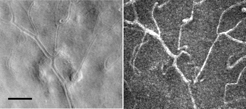
Adaptive Optics (AO) retinal imaging has provided revolutionary tools to scientists and clinicians for studying retinal structure and function in the living eye. From animal models to clinical patients, AO imaging is changing the way scientists are approaching the study of the retina. By providing cellular and subcellular details without the need for histology, it is now possible to perform large scale studies as well as to understand how an individual retina changes over time. Because AO retinal imaging is non-invasive and when performed with near-IR wavelengths both safe and easily tolerated by patients, it holds promise for being incorporated into clinical trials providing cell specific approaches to monitoring diseases and therapeutic interventions. AO is being used to enhance the ability of OCT, fluorescence imaging, and reflectance imaging. By incorporating imaging that is sensitive to differences in the scattering properties of retinal tissue, it is especially sensitive to disease, which can drastically impact retinal tissue properties. This review examines human AO retinal imaging with a concentration on the use of the Adaptive Optics Scanning Laser Ophthalmoscope (AOSLO). It first covers the background and the overall approaches to human AO retinal imaging, and the technology involved, and then concentrates on using AO retinal imaging to study the structure and function of the retina.

Keywords: Blood flow; Imaging; Ophthalmoscopy; Photoreceptors; Retina; Retinal degenerations; Vascular disease.


Figures

Fig. 1.
Fig. 1.
An example of AOSLO image of the retina. A: A small region of parafoveal retina imaged with an AOSLO that has been adjusted for best focus but without adaptive optics compensation of high order aberrations. While some cones are visible as low contrast spots, not all are visible. B: The same region of the retina, with the AO control loop activated, showing all cones within this region. C: A montage of foveal confocal AOSLO images showing the cone mosaic across the central retina. The location of highest cone density is marked with an asterisk. All scale bars 50 μm.
Fig. 2.
Fig. 2.
A schematic comparison of flood illuminated and scanning confocal imaging systems. Left: In a flood illuminated system the entire region of interest of the retina is illuminated simultaneously (blue). The illumination light enters the eye through a relatively small region of the pupil. Light returning from the retina (pink) is passed through the optical system and imaged onto an areas sensor, such as a camera, optically conjugate to the plane of focus. Light scattered within the eye (not shown), falls on the camera, but is out of focus. Left Inset: Resolution for a diffraction limited imaging system is controlled by the angle (θ) the pupil subtends with respect to the retina, that is the sine of the radius of the pupil divided by the distance between the retina and pupil. Right: a confocal imaging system illuminates the retina by scanning a small spot of light focused onto the retina (inset). Light returning from the sample is then separated from the input light by a beam splitter, and refocused onto a confocal aperture. Because the confocal aperture is optically conjugate to the point of focus of the imaging beam, all of the light backscattered from the focus of the beam is re-imaged at the aperture and therefore passes through the aperture to the detector, generating a series of signals, each corresponding to a particular retinal location, and thus forms an image. Light scattered from objects that lie elsewhere (blue) is not focused at the aperture and does not contribute to the image. Thus a confocal system provides “optical sectioning” and increased contrast, being insensitive to out of focus light. Right Inset: To create an image of the entire region of interest the imaging system scans the retina, building up a view of the retina one point at a time.
Fig. 3.
Fig. 3.
Schematic diagram of the three main classes of AO systems. Left: hardware based AO (HAO system). For HAO there are two primary components, the imaging system and the wavefront correction system. In the HAO system the control loop includes a wavefront sensor, such as the Shack Hartman sensor, which measures the optical aberrations. These measurement are fed back to the wavefront shaping system, such as by altering the shape of a deformable mirror, which in turn alters the optical properties of the imaging and detecting channels as both the imaging beam and the detecting beam are affected by the wavefront shaping. The wavefront is then measured again, and another correction is made, with measuring and corrections repeating until the noise level of the measuring and correcting system are reached. Center: Sensorless AO (SAO). For SAO the wavefront sensor is eliminated and the properties of the image itself are used to provide an input to the control loop. The system computes an image quality metric for each loop of the control algorithm, and the wavefront shaping device is then altered, the result measured, and the loop iterates until it converges on a diffraction limited image. Right: Computational AO (CAO) goes even further in reducing the hardware by omitting the wavefront shaping device and the need to iterate a control algorithm while imaging. The data are collected using a phase sensitive measurement, and an algorithm then computationally converges on the required wavefront correction. This correction is then implemented as a digital filter applied to the input image.
Fig. 4.
Fig. 4.
Operation of the Shack-Hartmann (SH) sensor. A: for a light source located at the focus of a perfect, diffraction limited lens, light will emerge from the lens perfectly collimated (all rays parallel). B: These parallel rays then come to an array of small lenses (lenslets) with an imaging sensor located one focal length behind the lenslets. Because the wavefront at each lenslet is flat, and orthogonal to the optical axis of the lens, each lenslet will produce a diffraction limited spot along its own optical axis on the image sensor. C: For a real eye, if a spot is imaged on the retina, light from that spot does not emerge from the eye perfectly collimated, but instead the rays vary with pupil location. This produces a non-flat wavefront. D: When this wavefront is reimaged at the SH sensor, each lenslet then has a wavefront impinging on it from slightly different angles. Since a lens produces an image offset from the optical axis at an angle proportional to the input angle, this means that a series of spots will be formed on the sensor, where the displacement of the spot relative to the optical axis of the lens will be proportional to the slope of the wavefront seen by each lenslet.
Fig. 5.
Fig. 5.
Comparison of bright field and dark field imaging in microscopy and in retinal imaging. A: dark field transmission microscopy passes light through the target and objects are typically seen as decrements on a bright background. B: In dark field microscopy an annular stop blocks undeviated light but passes more deviated light, showing objects as bright on a dark background. C: An SLO works in epi-illumination, which means the illumination and sensing are both from the same side, in this case the human pupil. Here some light is forward scattered at high angles from the focal spot (red triangle) D: Confocal imaging with an SLO puts an aperture conjugate to the focused spot of the illumination, and thus only light that is coming from the focal volume is detected. E: In multiply scattered light imaging, the directly backscattered light is blocked by a stop and the aperture, here a displaced aperture, lets through light that has been forward scattered and then scattered a second time and returned through the aperture, similar to the dark field microscopy and thus multiply scattered light imaging can reveal translucent structures.
Fig. 6.
Fig. 6.
Retinal capillary remodeling and edema in a diabetic subject showing capabilities of different imaging modes used simultaneously. A: Retinal capillaries imaged using a confocal aperture with a 2x Airy disc (AD) pinhole. B: The same capillaries imaged using an offset aperture with a 10x AD aperture displaced 6 AD diameters. C: A perfusion map of the same capillaries. The perfusion map is computed from variations in intensity that arise from blood flowing through the capillaries. D: Half annulus image. Complement of the half annulus image (Sapoznik et al, 2018). F: The same capillaries in a contrast image computed as (D-E)/(D+E). The contrast of various features changes with the mode of imaging. A capillary loop and surrounding area of edema (grey arrows) are least visible in the confocal image and most visible in the vascular map (C) or offset aperture image (B). A larger area of edema (white arrow) is readily detected in most of the images, but shows different characteristics, across imaging modes. Vascular walls are visible in capillaries, and best seen in the contrast image (F: black arrow). Scale bar 100 μm.
Fig. 7.
Fig. 7.
Rapid Acquisition of sequential AOSLO images using beam steering to build a montage that maps the cones from +10 to −10 degrees in a 34 year old male. Images were collected as averages of 100 frames per grab. After each video sequence the system displaced the beam by 1 degree, and acquired the next, generating a 22×3 degree montage in about 12 min. After this a more densely sampled foveal array was collected at a digital resolution of 0.67 mm/pixel, again using an automated sequence taking about 3 minutes. A: Image quality is good into the temporal retina out to 10 degrees (box and panel E), and in the foveal center (box and panel D), as well as at 10 degrees in the nasal retina (C). Image quality decreases somewhat at the edge of the steering field (box and panel B) requiring moving the fixation target. Scale bars 50 μm.
Fig. 8.
Fig. 8.
Left: Montage of AOSLO cone photoreceptor images obtained over the central 1 mm of the retina in a normal subject. Right: Cone density plot, based on semi-automated identification of all cones in the image on the left. The density plot shows the horizontal/vertical asymmetry in cone packing density. Scale bar 250 μm.
Fig. 9
Fig. 9
Fourier analysis of cone frequency. A: an expanded view of the central region of figure 6 (scale bar 25) μm. Spatial frequency analysis analyzes a small regions of cones (white box), B: the selected region, multiplied by a spatial mask to avoid windowing artifacts C: the log magnitude of the Fourier transform of the area. D: the radially averaged log power of the Fourier transform showing the spatial frequency of the cone sampling array (Yellot’s ring, arrow). Scale bar 50 μm.
Fig. 10.
Fig. 10.
Comparison of Voronoi analysis and local spatial average analysis. A: a group of cones. B: Voronoi analysis of the data in A. Here hexagonal regions are marked in a mid-gray. This region has a high degree of hexagonality (78% of regions). C: Local cone spatial average of the same region. This represents the average surround of all the cones in the analyzed region. Here we see that the array is uniform out to several rings of nearest neighbors. D: The radial average of the local cone spatial average, showing the regularity of the sampling array over this region. Scale bar 10 μm.
Fig. 11.
Fig. 11.
Example of rod imaging and cone imaging with both confocal and multiply scattered light. A: Photoreceptor image in the near periphery of the retina. Here the cones (larger cells) are surrounded with smaller rods. B: Confocal image with a 2 degree field. C: The same region, imaged simultaneously with B, but using a half annular aperture for multiply scattered light detection. For the multiply scattered light image the cone inner segments are clearly visible as dots with a bright crescent opposite to the side of the half annular aperture. Note that while for the confocal image only cones are visible, for the multiply scattered light image the background of the image shows a strong contribution from choroidal scattering, most likely the site of the majority of second scattering events, when focused on the cones, with the dark regions representing the choroidal blood vessels. Scale bar in A 25 μm. Scale bars in B and C 100 μm.
Fig. 12.
Fig. 12.
Cones are seen extending out onto atrophic regions or the retina. A: An image of the nerve fiber layer at the edge of the optic nerve. B: The same retinal region as in A but now focused at the cones, showing cones extending onto the peripapillary crescent. Data replotted from Chui et al, (Chui et al., 2011) C: Cones in an outer retinal tubulation showing the bright cones extending out across a regions of atrophy. Data replotted from King et al. (King et al., 2017). Scale bars in A and B 100 μm. Scale bar in C 25 μm.
Fig. 13.
Fig. 13.
A comparison of some of the structural changes in diabetic retinopathy. While typically we grade the retina on inner retinal changes, there are also changes occurring deeper. A: An example of a region of multiple vascular anomalies, including looping and doubling of capillaries (white arrows) and non-perfused capillaries (black arrows) in early diabetic retinopathy. B. A region of microcystic changes (dark regions), not visible at normal magnification but seen with AO retinal imaging. There is a small capillary loop present at the edge of the foveal avascular zone. C: The localized regions of dark cones apparent in about 25% of diabetic patients. The failure to guide light in consistent with an early stage of failure of the outer blood retinal barrier. The large region in the top center has been present for 3 years (Sawides et al., 2017b). All images have been scaled for display, with the cones imaging using a logarithmic reflectance scale. Scale bars for all panels 100 μm.
Fig. 14.
Fig. 14.
Foveal avascular zone maps from a normal subject obtained using clinical OCT-A and adaptive optics imaging. A: Heidelberg Spectralis OCTA imaging. B: Split image with the left region being a magnification of the central fovea from panel A and the right being the vascular map obtained using a laboratory based AOSLO using offset pinhole detection. In both cases the superficial vascular plexus was used. Almost all vessels are visible using the OCT-A, however the exact locations and some vessels which are within 20 um are not well resolved with the clinical instrumentation. Scale bars are 200 μm.
Fig. 15.
Fig. 15.
Detection of non-perfused capillaries with half annulus contrast image (Sapoznik et al, 2018). Left: AOSLO image of a diabetic subject. Right: the vascular map obtained simultaneously. White arrows point to capillaries visible in the structural image but lacking flow. Scale bar 100 μm.
Fig. 16.
Fig. 16.
Combining perfusion maps with time resolved velocity measurements. Left: An example of a region of the superior vascular plexus where vessels determined by the motion of red blood cells. Arrows indicate the direction of flow from the arteriole (bottom right) towards the venule (top left). Right: Blood velocity measurement in two venules (indicated by the color of the arrows). The pulsatility and velocity in these two venules are similar, even though they are at different levels of the vascular tree, presumably because the increase in size of the venule is sufficient to carry the converging flows from the capillaries. Scale bar 50 μm.
Fig. 17.
Fig. 17.
Left: The walls of the retinal vessels and even the cellular components of the walls can be visualized with multiply scattered light imaging. Right image sequence: With confocal imaging and many clinical instruments however a bright stripe is often seen in the center of the arteries. This arterial reflex appears to be related to the velocity of red blood cells. As shown here the stripe varies during a single cardiac cycle starting at a velocity minimum (diastolic) on the left, through a peak at the systolic phase and back to a trough, as seen by the slope of individual red blood cells moving across the stabilized line (see discussion under blood flow). Scale bar 100 μm.
Fig. 18.
Fig. 18.
Examples of wall to lumen ration measurements using AOSLO measurements. An arteriole in a subject with hypertension measured using a split annular aperture. Arrows indicate the outer and inner edges of the walls. B: A set of measurements in control and hypertensive subjects showing the strong covariance between the vessel lumen (inner width) and the wall to lumen ratio. This covariance adds noise to clinical measurements unless matched samples are used. C: For normal subjects measuring the walls over the entire range of sizes available demonstrates that inner and outer arterial diameters are tightly controlled. The dashed line shows a slope of 1, so the deviation from that slope illustrates the relative increase in wall thickness with increasing vessel size. Comparing patient results to the linear fit (solid line) normalize the data, increasing the statistical power of measurements (Hillard et al, 2017, data replotted from Hillard et al. 2017). D: The walls of diabetic subject are also thickened and similar techniques can be used to reduce the data but in addition they often show variability in the mural cells (black arrows). Scale bars 100 μm.
Fig.19.
Fig.19.
Comparison of standard fundus view and AO retinal imaging of a diabetic retina. Left: Heidelberg near infrared SLO image of the retina of a 36 year old diabetic female showing a few local changes. Right: the highly magnified AOSLO image of a small region of the retina shown in the white box. Here a single region shows abnormal vessel walls (open black arrows), capillary loops and tangles (black arrows), and non-perfused capillaries (white arrows). Also shown are areas of wall thickening (white arrows with black outlines), which seems to co-locate with other retinal changes. Scale bar 100 μm.
Fig. 20.
Fig. 20.
Examples of cotton wool spot (CWS) eleven days after the initial vascular event. Top: the extent of the damage can be seen coursing from the optic nerve, up to the CWS (small box A), and out across the retina. Scale bar 500 μm. A: At the location of the CWS cytoid bodies can be seen (white arrow). Further from the CWS fields of small round spots were seen (boxes B and C, black arrows). These are presumed apoptotic ganglion cells and the fall solely along the nerve fiber bundles passing through the CWS and peaked about 9 days after the CWS spot occurred and then decreased in number to close to zero at 30 days after the insult. Scale bars for A, B and C 100 μm.
Fig. 21.
Fig. 21.
Example of changes in deep retinal images in a serous detachment of the retina. A: OCT b-scan of a patient with a serous detachment secondary to a nevus. The red box indicates the approximate locations of panels B and C. B: Offset aperture images focused on the cones, showing regularly arranged mosaic of inner segments. Offset aperture images focused on the RPE, showing an array of RPE cells. Scale bars: A, 200 μm, B and C, 50 μm.
Fig. 22.
Fig. 22.
Examples of putative glial cells near the optic disc. A: A series of Gunn dots imaged with a slightly offset aperture configuration. B: A more densely sampled image using a half annulus aperture, showing the cell outline and extensions to the nearby vein. C: A confocal image showing putative glial connections to a vessel. These types of connections are most common near the optic nerve. Scale bars 50 μm.
Fig. 23.
Fig. 23.
An example of an AOSLO being used to present a stimulus directly to the retina by modulating the light beam. Here an AOM is modulating the light, while the retina is being stabilized using a strip based image alignment technique (Arathorn et al., 2007).
Fig. 24.
Fig. 24.
AOSLO image frames collected at 29 Hz using an offset aperture. A: reflectance image showing two regions where there are continuous sequences of red blood cells moving through two capillaries (white arrows). B: A single frame subregion of A, showing both a group of red blood cells in rouleaux (black arrow) followed by a plasma gap (white arrow). C and D: Same region as in B, but now showing two sequential frames, computed by calculating the difference from the mean of 100 frames (in standard deviations). Here we can see that the motion of the rouleaux is readily measured by its change in position. Scale bars are 50 μm.
Fig. 25.
Fig. 25.
AOSLO image in line scan mode. Here the slow scan position is controlled by a temporally changing voltage. Left: This voltage is programmed such that over part of the frame it follows a normal ramp (time period a), for the second part of the frame the slow scan is stopped (b). Right, the resulting slow scan pattern creates a retinal image where the top (a) is a normal view of the retina, and this allows detection of gross eye motions. The bottom portion of the image (b in right side image) has a stationary slow scan, and during this time period generates an xt image (x-position vs time) image (b). For a blood vessel that crosses the line scan, motion of particles in the x-direction create slanted lines, where the slope of the line is the horizontal component of the cell velocity. Retinal features that are not moving appear as vertical lines during this time period, allowing correction of horizontal eye motion, but not vertical motions within the frame. Scale bar 100 μm.
Fig. 26.
Fig. 26.
AOSLO blood flow imaging using dual scanning. A: single frame offset aperture image from channel 1. B: Single frame from channel 2, captured simultaneously with A. The two channels are slightly displaced from each other at the retina, resulting in a given location being imaged at two slightly different times. In this case about 5 msec apart. C: For every pixel a z-score is computed at each point in time for the likelihood of a change. Here the z-score for the frame in A is shown. The white arrows show a group of erythrocytes in a capillary. D: The z-scores for B. E: For each detected cell in a given capillary, a region of interest is extracted with the cell at the center. These regions of interests are averaged, producing image E. F: Average of the same ROI’s calculated for channel 2. Here the average shows a bright cell that is not centered, but is rather displaced from the center, with the amount of displacement indicating the difference traveled in 5 msec. G: The average velocity for the cell, averaged over 3 consecutive video frames, for the entire 3 second video, showing the pulsatile change in velocity which is at the same frequency as the cardiac cycle. Scale bars in A and B, 50 μm.

References

    1. Adie SG, Graf BW, Ahmad A, Carney PS, Boppart SA, 2012. Computational adaptive optics for broadband optical interferometric tomography of biological tissue. Proceedings of the National Academy of Sciences of the United States of America 109, 7175–7180. - PMC - PubMed
    1. Agabiti-Rosei E, Rizzoni D, 2017. Microvascular structure as a prognostically relevant endpoint. Journal of Hypertension 35, 914–921. - PubMed
    1. Antonetti DA, Barber AJ, Bronson SK, Freeman WM, Gardner TW, Jefferson LS, Kester M, Kimball SR, Krady JK, LaNoue KF, Norbury CC, Quinn PG, Sandirasegarane L, Simpson IA, Grp JDRC, 2006. Diabetic retinopathy - Seeing beyond glucose-induced microvascular disease. Diabetes 55, 2401–2411. - PubMed
    1. Arathorn DW, Yang Q, Vogel CR, Zhang Y, Tiruveedhula P, Roorda A, 2007. Retinally stabilized cone-targeted stimulus delivery. Opt Express 15, 13731–13744. - PubMed
    1. Arichika S, Uji A, Ooto S, Miyamoto K, Yoshimura N, 2014. Adaptive Optics-Assisted Identification of Preferential Erythrocyte Aggregate Pathways in the Human Retinal Microvasculature. Plos One 9, e89679. - PMC - PubMed

Publication types

MeSH terms