

A morphological classification for vocal fold leukoplakia
- PMID: 30166121
- PMCID: PMC9443019
- DOI: 10.1016/j.bjorl.2018.04.014
A morphological classification for vocal fold leukoplakia
Abstract
Introduction: There is still no general method for discriminating between benign and malignant leukoplakia and identifying vocal fold leukoplakia.
Objective: To evaluate the reliability of a morphological classification and the correlation between morphological types and pathological grades of vocal fold leukoplakia.
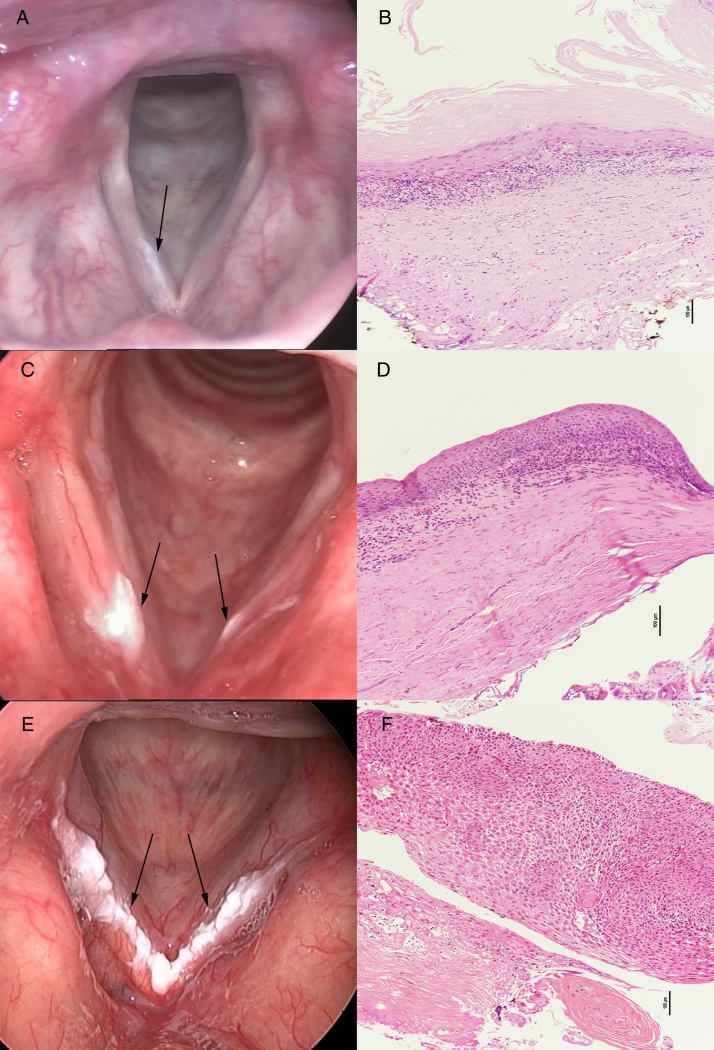
Methods: A total of 375 patients with vocal fold leukoplakia between 2009 and 2015 were retrospectively reviewed. Two observers divided the vocal fold leukoplakia into flat and smooth, elevated and smooth, and rough type on the basis of morphological appearance. The inter-observer reliability was evaluated and the results of classification from both observers were compared with final pathological grades. Clinical characteristics between low risk and high risk group were also analyzed.
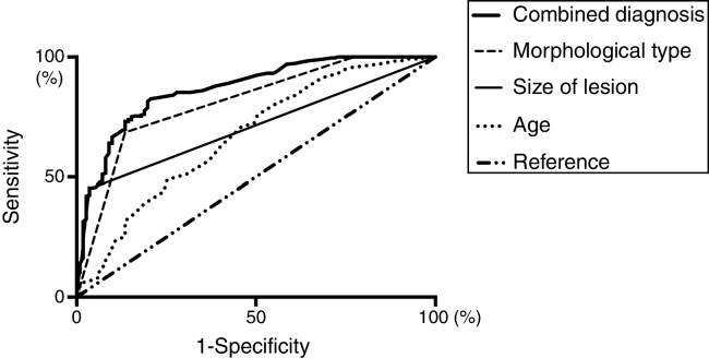
Results: The percentage inter-observer agreement of the morphological classification was 78.7% (κ=0.615, p<0.001). In the results from both observers, the morphological types were significantly correlated with the pathological grades (p1<0.001, p2<0.001, Kruskal-Wallis test; r1=0.646, p1<0.001, r2=0.539, p2<0.001, Spearman Correlation Analysis). Multivariate analysis showed patient's age (p=0.018), the size of lesion (p<0.001), and morphological type (p<0.001) were significantly different between low risk group and high risk group. Combined receiver operating characteristic curve analysis of significant parameters revealed an area under the receiver operating characteristic curve of 0.863 (95% CI 0.823-0.903, p<0.001).
Conclusions: The proposed morphological classification of vocal fold leukoplakia was consistent between observers and morphological types correlated with pathological grades. Patient's age, the size of lesion, and morphological type might enable risk stratification and provide treatment guidelines for vocal fold leukoplakia.
Introdução: Ainda não há um método universal estabelecido para diferenciar entre a leucoplasia benigna e maligna ou identificar as leucoplasias das pregas vocais.
Objetivo: Avaliar a confiabilidade de uma classificação morfológica e a correlação entre os tipos morfológicos e os graus histopatológicos das leucoplasias de pregas vocais.
Método: Os registros de 375 pacientes com leucoplasia da prega vocal assistidos entre 2009 e 2015 foram revisados retrospectivamente. Dois observadores dividiram a leucoplasia da prega vocal entre tipo plano e liso, elevado e liso, e rugoso, com base na aparência morfológica. A confiabilidade interobservador foi avaliada e os resultados de classificação de ambos os observadores foram comparados com os graus histopatológicos finais. As características clínicas entre os grupos de baixo risco e alto risco também foram analisadas.
Resultados: A porcentagem da concordância inter-observador da classificação morfológica foi de 78,7% (κ = 0,615, p < 0,001). Nos resultados de ambos os observadores, os tipos morfológicos correlacionaram-se significativamente com os graus histopatológicos (p1 < 0,001, p2 < 0,001, teste de Kruskal-Wallis; r1 = 0,646, p1 < 0,001, r2 = 0,539, p2 < 0,001, análise de correlação de Spearman). A análise multivariada mostrou que a idade do paciente (p = 0,018), o tamanho da lesão (p < 0,001) e o tipo morfológico (p < 0,001) foram significativamente diferentes entre o grupo de baixo risco e o de alto risco. A análise da curva ROC (Receiver Operating Characteristic) combinada de parâmetros significativos revelou uma área sob a curva de 0,863 (IC 95%: 0,823 ± 0,903, p < 0,001).
Conclusões: A classificação morfológica proposta para leucoplasia de prega vocal foi consistente entre observadores e os tipos morfológicos correlacionaram-se com os graus histopatológicos. A idade do paciente, o tamanho da lesão e o tipo morfológico podem permitir a estratificação de risco e fornecem diretrizes de tratamento para a leucoplasia da prega vocal.
Keywords: Displasia; Dysplasia; Leucoplasia; Leukoplakia; Morfológico; Morphological; Pathological; Patológico; Prega vocal; Vocal fold.
Copyright © 2019 Associação Brasileira de Otorrinolaringologia e Cirurgia Cérvico-Facial. Published by Elsevier Editora Ltda. All rights reserved.
Figures
References
-
- Panwar A., Lindau R., 3rd, Wieland A. Management of premalignant lesions of the larynx. Expert Rev Anticancer Ther. 2013;13:1045–1051. - PubMed
-
- Thompson L. World Health Organization classification of tumours: pathology and genetics of head and neck tumours. Ear Nose Throat J. 2006;85:74. - PubMed
-
- Gale N., Gnepp D.R., Poljak M., Strojan P., Cardesa A., Helliwell T., et al. Laryngeal squamous intraepithelial lesions: an updated review on etiology, classification, molecular changes, and treatment. Adv Anat Pathol. 2016;23:84–91. - PubMed
MeSH terms
LinkOut - more resources
Full Text Sources
Other Literature Sources
