. 2018 Nov;17(11):2365-2376.
doi: 10.1158/1535-7163.MCT-18-0176. Epub 2018 Aug 30.

Targeted Inhibition of ULK1 Promotes Apoptosis and Suppresses Tumor Growth and Metastasis in Neuroblastoma

Affiliations

Targeted Inhibition of ULK1 Promotes Apoptosis and Suppresses Tumor Growth and Metastasis in Neuroblastoma

Christopher M Dower et al. Mol Cancer Ther. 2018 Nov.

Abstract

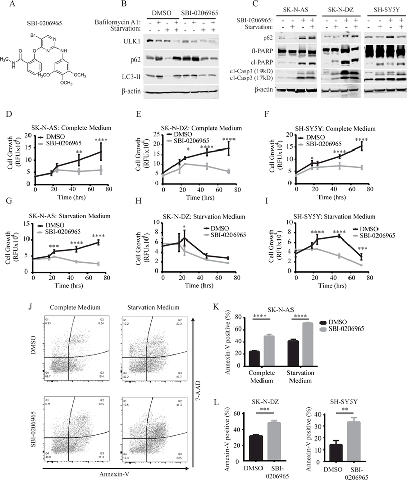
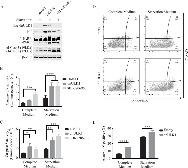
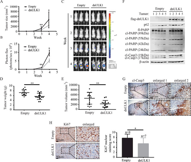
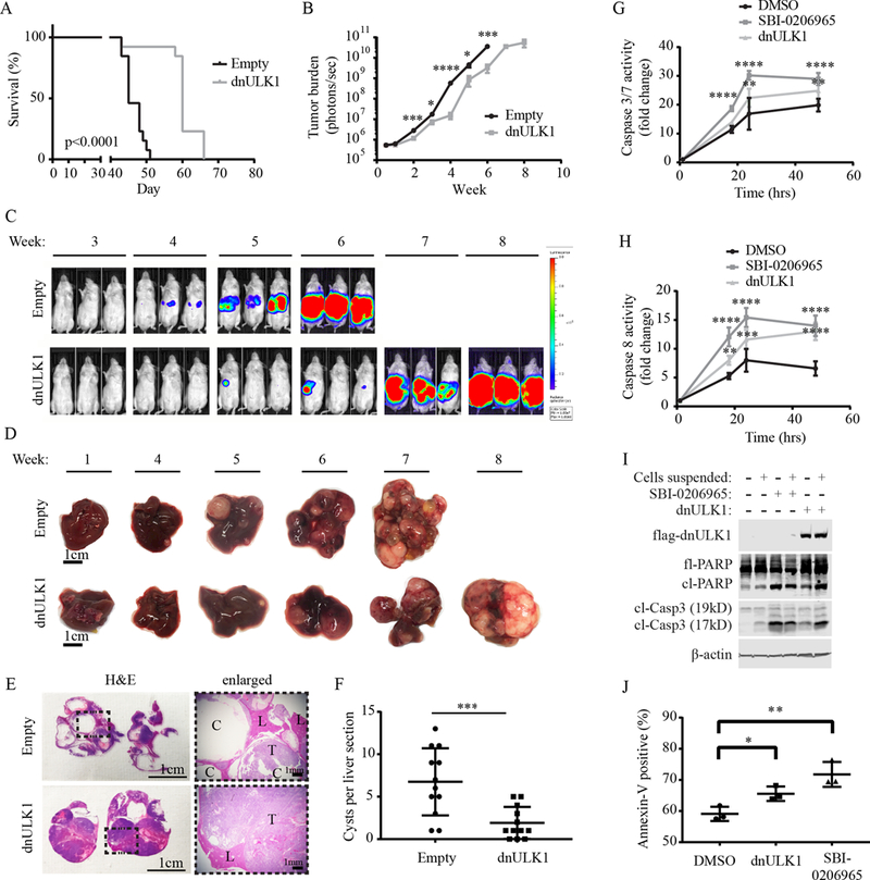
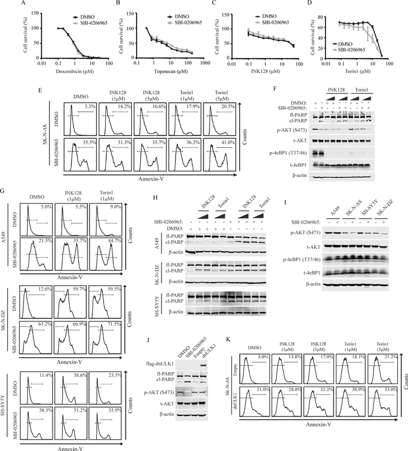
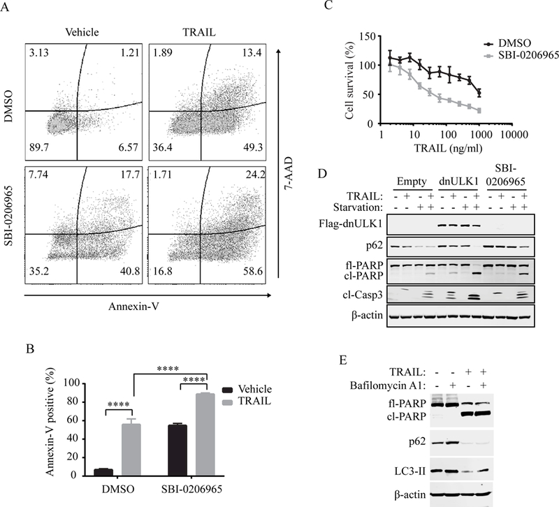
Neuroblastoma is the most common extracranial solid malignancy in the pediatric population, accounting for over 9% of all cancer-related deaths in children. Autophagy is a cell self-protective mechanism that promotes tumor cell growth and survival, making it an attractive target for treating cancer. However, the role of autophagy in neuroblastoma tumor growth and metastasis is largely undefined. Here we demonstrate that targeted inhibition of an essential autophagy kinase, unc-51 like autophagy kinase 1 (ULK1), with a recently developed small-molecule inhibitor of ULK1, SBI-0206965, significantly reduces cell growth and promotes apoptosis in SK-N-AS, SH-SY5Y, and SK-N-DZ neuroblastoma cell lines. Furthermore, inhibition of ULK1 by a dominant-negative mutant of ULK1 (dnULK1K46N) significantly reduces growth and metastatic disease and prolongs survival of mice bearing SK-N-AS xenograft tumors. We also show that SBI-0206965 sensitizes SK-N-AS cells to TRAIL treatment, but not to mTOR inhibitors (INK128, Torin1) or topoisomerase inhibitors (doxorubicin, topotecan). Collectively, these findings demonstrate that ULK1 is a viable drug target and suggest that inhibitors of ULK1 may provide a novel therapeutic option for the treatment of neuroblastoma. Mol Cancer Ther; 17(11); 2365-76. ©2018 AACR.


Figures

Figure 1.
Figure 1.. SBI-0206965 reduces cell growth and promotes apoptosis in neuroblastoma cell lines.
(A) Chemical structure of SBI-0206965. (B) Autophagic flux assay assessing the functional effects of SBI-0206965 on autophagy and ULK1 protein levels. (C)Western blot analysis of autophagy (p62) and apoptosis (cleaved-PARP and cleaved-caspase 3) markers after treatment with 10¼M SBI-0206965 for 48 hours in SK-N-AS and 24 hours in SK-N-DZ and SH-SY5Y cells. (D-I) Cell growth as assessed by Prestoblue in the indicated neuroblastoma cell lines treated with either DMSO or 10¼M SBI-0206965 over 72 hours in complete medium (D-F) or starvation medium (G-I). (J-K) Annexin-V/7-AAD flow cytometry analysis of SK-N-AS cells treated with 10¼M SBI-0206965 or DMSO for 48 hours in complete medium or starvation medium. (L) Annexin-V/7-AAD flow cytometry analysis of SK-N-DZ and SH-SY5Y cells treated with DMSO or 10¼M SBI-0206965 for 24 hours in starvation medium. *p<0.05, **p<0.01, ***p<0.001, p<0.0001.
Figure 2.
Figure 2.. Inhibition of ULK1 by dnULK1 promotes apoptosis in SK-N-AS cells.
(A) Western blot analysis of SK-N-AS cells expressing dnULK1K46N or treated with 10¼M SBI-0206965 or DMSO for 48 hours in complete or starvation medium. (B-C) Assessment of caspase-3/7 activity (B) or caspaspe-8 activity (C) of SK-N-AS cells expressing dnULK1K46N construct or treated with 10¼M SBI-0206965 or DMSO for 24 hours in complete or starvation medium. (D-E) Annexin-V/7-AAD flow cytometry assay of SK-N-AS cells expressing dnULK1K46N or empty-vector control in complete medium or starvation medium for 48 hours. *p<0.05, **p<0.01, ***p<0.001, p<0.0001.
Figure 3.
Figure 3.. Inhibition of ULK1 by dnULK1 reduces SK-N-AS xenograft tumor growth and promotes apoptosis.
(A) Tumor volume of SK-N-AS xenografts expressing either dnULK1 or empty-vector over 4 weeks (mean ± SD, n=16 mice per group). (B) Quantification of luciferase intensity (photons/sec) in dnULK1 and empty-vector tumors (mean ± SEM, n=16 mice per group). (C) Representative images of luciferase expression from primary xenograft tumors over a 4-week time period. (D) Ex vivo analysis of xenograft tumor weight (mean ± SD). (E) Ex vivo analysis of xenograft tumor volume (mean ± SD). (F) Western blot analysis of xenograft tumors (n=5 tumors per group). (G) Representative image of immunohistochemical staining for cleaved caspase-3 of primary xenograft tumors. (H) Quantification and representative image of immunohistochemical staining for Ki67 of primary xenograft tumors (mean ± SD). *p<0.05, **p<0.01
Figure 4.
Figure 4.. Inhibition of ULK1 by dnULK1 promotes survival and reduces metastatic tumor growth.
(A) Kaplan-Meier survival curve of mice injected via the tail vein with dnULK1-expressing SK-N-AS cells compared to empty-vector control. (B) Quantification of metastatic tumor growth via luciferase expression (mean ± SEM, n = 13). (C) Representative images of luciferase expression in SK-N-AS xenografts. (D) Representative images of tumor-bearing livers over an 8-week time period. (E) Representative images of H&E staining of tumor-bearing liver sections from empty-vector mice at week 7 and dnULK1 mice at week 8. (C=cyst, L=liver tissue, T=tumor tissue). (F) Quantification of cysts in empty-vector and dnULK1 liver sections. (G) Caspase 3/7 activity in suspended SK-N-AS cells expressing dnULK1 or treated with 10¼M SBI-0206965 or DMSO over 48 hours. (H) Caspase 8 activity in suspended SK-N-AS cells expressing dnULK1 or treated with 10¼M SBI-0206965 or DMSO over 48 hours. (I) Western blot analysis of adherent and suspended SK-N-AS cells expressing dnULK1 or treated with 10¼M SBI-0206965 or DMSO for 48 hours. (J) Assessment of annexin-V staining by flow cytometry in suspended SK-N-AS cells expressing dnULK1 or treated with 10¼M SBI-0206965 or DMSO for 48 hours. *p<0.05, **p<0.01, ***p<0.001, ****p<0.0001.
Figure 5.
Figure 5.. SBI-0206965 does not affect the cytotoxicity of topoisomerase or mTOR inhibitors in neuroblastoma cells.
(A-D) Survival curve measured by Prestoblue of SK-N-AS cells co-treated with increasing concentrations of doxorubicin (A), topotecan (B), INK128 (C), or Torin1 (D) and 10μM SBI-0206965 or DMSO vehicle for 24 hours. (E) Annexin-V flow cytometry analysis of SK-N-AS cells treated with 10μM SBI-0206965 and 5¼M INK128 or Torin1 alone or in combination for 48 hours. (F) Western blot analysis of SK-N-AS cells treated with 10μM SBI-0206965 and INK128 or Torin1 at 1¼M and 5¼M alone or in combination for 48 hours. (G) Annexin-V flow cytometry analysis of A549, SH-SY5Y and SK-N-DZ cells treated with 10μM SBI-0206965 and 1¼M INK128 or Torin1 alone or in combination. A549 cell were treated for 48 hours. SH-SY5Y and SK-N-DZ cells were treated for 24 hours. (H) Western blot analysis of A549, SH-SY5Y and SK-N-DZ cells treated with 10μM SBI-0206965 and INK128 or Torin1 at 1¼M and 5¼M alone or in combination. A549 cell were treated for 48 hours. SH-SY5Y and SK-N-DZ cells were treated for 24 hours. (I) Western blot analysis assessing mTORC1/2 downstream signaling in A549, SK-N-AS, SH-SY5Y and SK-N-DZ treated with SBI-0206965 or DMSO control for 24 hours. (J) Western blot analysis comparing phosphorylated AKT(S473) levels in SK-N-AS cells expressing dnULK1K46N or treated with SBI-0206965 for 48 hours and empty-vector or DMSO control. (K) Annexin-V flow cytometry analysis of SK-N-AS expressing dnULK1 or empty-vector treated with INK128 or Torin1 at 1¼M and 5¼M for 48 hours.
Figure 6.
Figure 6.. SBI-0206965 sensitizes SK-N-AS cells to TRAIL treatment.
(A-B) Annexin-V/7-AAD flow cytometry analysis of SK-N-AS cells. Cells were treated with 10¼M SBI-0206965 or DMSO alone for 44 hours, followed by the addition of 100ng/ml TRAIL or vehicle for 4 hours, for a total treatment time of 48 hours. (C) Survival curve measured by Prestoblue of SK-N-AS cells co-treated with increasing concentrations of TRAIL and 10¼M SBI-0206965 or DMSO for 24 hours. (D) Western blot analysis of SK-N-AS cells expressing empty-vector or dnULK1 or treated with 10¼M SBI-0206965 and 100ng/ml TRAIL alone or in combination for 4 hours in normal or starvation medium. ****p<0.0001. (E) Western blot analysis of autophagy markers in SK-N-AS cells treated with TRAIL or vehicle control for 6 hours.

References

    1. Irwin MS, Park JR. Neuroblastoma: paradigm for precision medicine. Pediatr Clin North Am. 2015;62:225–56. - PubMed
    1. Matthay KK, Maris JM, Schleiermacher G, Nakagawara A, Mackall CL, Diller L, et al. Neuroblastoma. Nat Rev Dis Primer. 2016;2:16078. - PubMed
    1. Vo KT, Matthay KK, Neuhaus J, London WB, Hero B, Ambros PF, et al. Clinical, Biologic, and Prognostic Differences on the Basis of Primary Tumor Site in Neuroblastoma: A Report From the International Neuroblastoma Risk Group Project. J Clin Oncol. 2014;32:3169–76. - PMC - PubMed
    1. Matthay KK, Villablanca JG, Seeger RC, Stram DO, Harris RE, Ramsay NK, et al. Treatment of high-risk neuroblastoma with intensive chemotherapy, radiotherapy, autologous bone marrow transplantation, and 13-cis-retinoic acid. Children’s Cancer Group. N Engl J Med. 1999;341:1165–73. - PubMed
    1. Pinto NR, Applebaum MA, Volchenboum SL, Matthay KK, London WB, Ambros PF, et al. Advances in Risk Classification and Treatment Strategies for Neuroblastoma. J Clin Oncol Off J Am Soc Clin Oncol. 2015;33:3008–17. - PMC - PubMed

Publication types

MeSH terms