. 2018 Oct 5;362(6410):86-91.
doi: 10.1126/science.aau1549. Epub 2018 Aug 30.

Gene editing restores dystrophin expression in a canine model of Duchenne muscular dystrophy

Affiliations

Gene editing restores dystrophin expression in a canine model of Duchenne muscular dystrophy

Leonela Amoasii et al. Science. .

Abstract

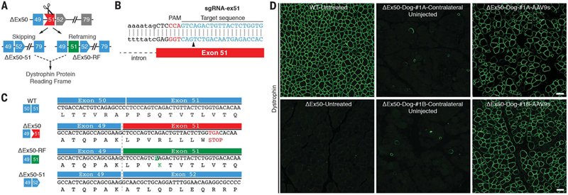
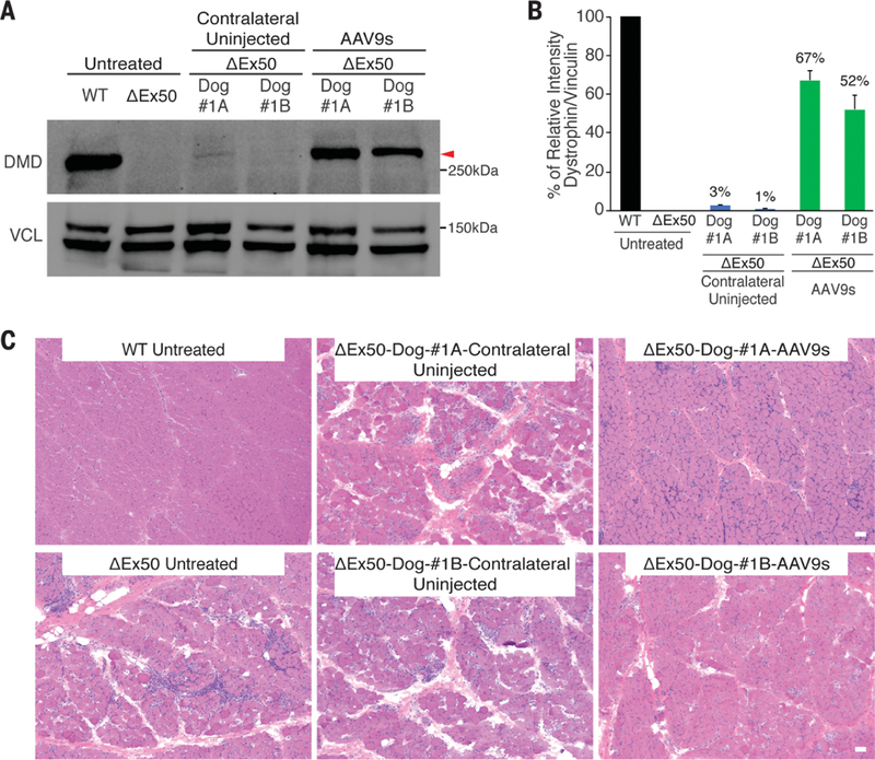
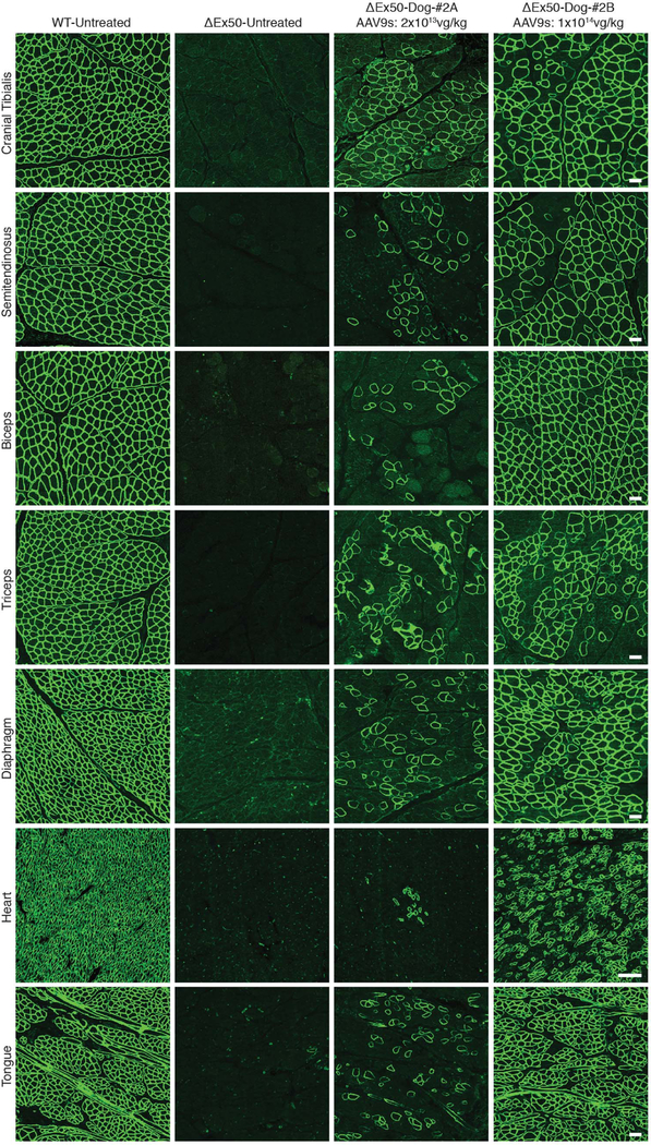
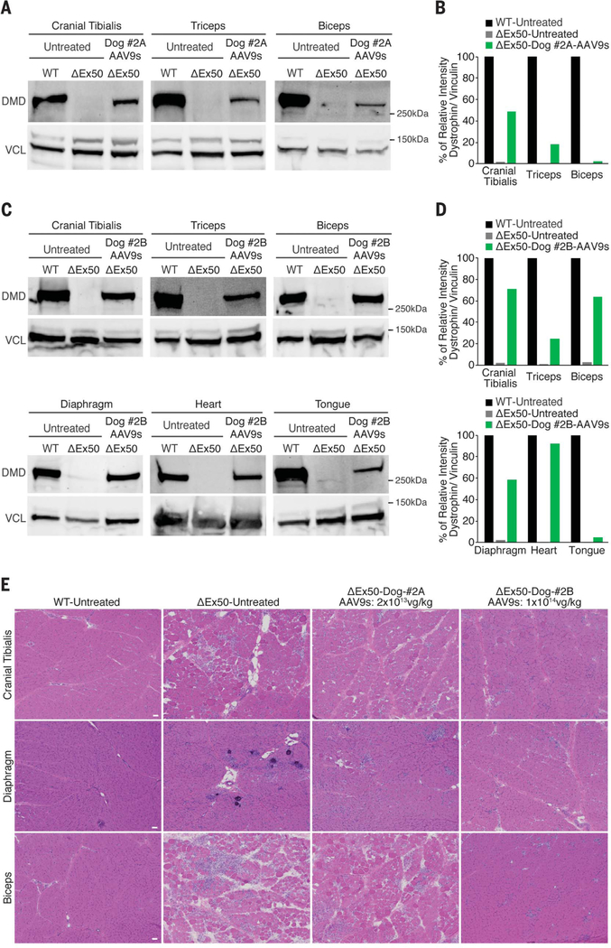
Mutations in the gene encoding dystrophin, a protein that maintains muscle integrity and function, cause Duchenne muscular dystrophy (DMD). The deltaE50-MD dog model of DMD harbors a mutation corresponding to a mutational "hotspot" in the human DMD gene. We used adeno-associated viruses to deliver CRISPR gene editing components to four dogs and examined dystrophin protein expression 6 weeks after intramuscular delivery (n = 2) or 8 weeks after systemic delivery (n = 2). After systemic delivery in skeletal muscle, dystrophin was restored to levels ranging from 3 to 90% of normal, depending on muscle type. In cardiac muscle, dystrophin levels in the dog receiving the highest dose reached 92% of normal. The treated dogs also showed improved muscle histology. These large-animal data support the concept that, with further development, gene editing approaches may prove clinically useful for the treatment of DMD.


Conflict of interest statement

Competing interests: L.A., R.B.-D., R.J.P., and E.N.O. are consultants for Exonics Therapeutics. L.A. and E.N.O. are co-inventors on a patent application (provisional filing number 62/442,606) related to the strategy presented in this study. The other authors declare no competing interests.

Figures

Fig. 1.
Fig. 1.. Single-cut CRISPR editing of canine exon 50 in vivo and in vitro.
(A) Scheme showing the CRISPR-Cas9–mediated genome editing approach to correct the reading frame in ΔEx50 dogs by reframing and skipping of exon 51. Gray exons are out of frame. (B) Illustration of sgRNA binding position and sequence for sgRNA-ex51. PAM sequence for sgRNA is indicated in red. Black arrowhead indicates the cleavage site. (C) Sequence of the RT-PCR products of the ΔEx50–51 lower band confirmed that exon 49 spliced directly to exon 52, excluding exon 51. Sequence of RT-PCR products of ΔEx50 reframed (ΔEx50-RF). (D) Cranial tibialis muscles of ΔEx50 dogs were injected with AAV9s encoding sgRNA-51 and Cas9 and analyzed 6 weeks later. Dystrophin immunohistochemistry staining of cranial tibialis muscle of wild-type (WT) dog untreated, ΔEx50 dog untreated, ΔEx50 dogs contralateral (uninjected) muscle, and ΔEx50 dogs injected with AAV9-Cas9 and AAV9-sgRNA-51 (referred to as ΔEx50-#1A-AAV9s and ΔEx50-#1B-AAV9s). Scale bar: 50 μm.
Fig. 2.
Fig. 2.. Dystrophin correction after intramuscular delivery of AAV9-encoded gene editing components.
(A) Western blot analysis of dystrophin (DMD) and vinculin (VCL) expression in cranial tibialis muscles 6 weeks after intramuscular injection in 2 dogs (#1A and #1B). (B) Quantification of dystrophin expression from blots after normalization to vinculin. (C) Histochemistry by H&E staining of cranial tibialis muscle from a WT dog, ΔEx50 dog untreated, ΔEx50 contralateral uninjected, and ΔEx50 dogs injected intramuscularly with AAV9-Cas9 and AAV9-sgRNA-51 (referred to as ΔEx50-Dog-#1A-AAV9s and ΔEx50-Dog-#1B-AAV9s). Scale bar: 50 µm.
Fig. 3.
Fig. 3.. Immunostaining of dystrophin after intravenous delivery of AAV9-encoded gene editing components.
Dystrophin immunohistochemistry staining of cranial tibialis, semitendinosus, biceps, triceps, diaphragm, heart, and tongue muscles of WT dog, untreated ΔEx50 dog, and ΔEx50 dogs injected systemically with AAV9-Cas9 and AAV9-sgRNA-51 at 2 × 10¹³vg/kg (total virus 4 × 10¹³ vg/kg, referred to as ΔEx50-Dog #2A-AAV9s) and 1 × 10¹⁴ vg/kg (total virus 2 × 10¹⁴ vg/kg, referred to as ΔEx50-Dog #2B-AAV9s) for each virus.
Fig. 4.
Fig. 4.. Western blot of dystrophin and muscle histology after intravenous delivery of AAV9-encoded gene editing components.
(A) Western blot analysis of dystrophin (DMD) and vinculin (VCL) of cranial tibialis, triceps, and biceps muscles of WT, untreated ΔEx50, and ΔEx50 injected with AAV9-Cas9 and AAV9-sgRNA-51 at 2 × 10¹³ vg/kg for each virus (total virus 4 × 10¹³ vg/kg, referred to as ΔEx50-Dog #2A-AAV9s). (B) Quantification of dystrophin expression from blots after normalization to vinculin. (C) Western blot analysis of DMD and VCL of cranial tibialis, triceps, biceps, diaphragm, heart, and tongue muscles of WT, untreated DEx50, and DEx50 injected with AAV9-Cas9 and AAV9-sgRNA-51 at 1 × 10¹⁴ vg/kg (total virus 2 × 10¹⁴ vg/kg, referred to as ΔEx50-Dog #2B-AAV9s). (D) Quantification of dystrophin expression from blots after normalization to vinculin. (E) H&E staining of cranial tibialis, diaphragm, and biceps muscles of WT, untreated ΔEx50, and ΔEx50 injected with AAV9-Cas9 and AAV9-sgRNA-51 at 2 × 10¹³ vg/kg for each virus (total virus 4 × 10¹³ vg/kg) and 1 × 10¹⁴vg/kg for each virus (total virus 2 × 10¹⁴ vg/kg). Scale bar: 50 μm.

Comment in

References

    1. O’Brien KF, Kunkel LM, Mol. Genet. Metab 74, 75–88 (2001). - PubMed
    1. Guiraud S et al., Annu. Rev. Genomics Hum. Genet 16, 281–308 (2015). - PubMed
    1. Campbell KP, Kahl SD, Nature 338, 259–262 (1989). - PubMed
    1. Ervasti JM, Ohlendieck K, Kahl SD, Gaver MG,Campbell KP, Nature 345, 315–319 (1990). - PubMed
    1. Muntoni F, Torelli S, Ferlini A, Lancet Neurol 2, 731–740 (2003). - PubMed

Publication types