. 2018 Aug 31;361(6405):eaao3048.
doi: 10.1126/science.aao3048.

Cancer mutations and targeted drugs can disrupt dynamic signal encoding by the Ras-Erk pathway

Affiliations

Cancer mutations and targeted drugs can disrupt dynamic signal encoding by the Ras-Erk pathway

L J Bugaj et al. Science. .

Abstract

The Ras-Erk (extracellular signal-regulated kinase) pathway encodes information in its dynamics; the duration and frequency of Erk activity can specify distinct cell fates. To enable dynamic encoding, temporal information must be accurately transmitted from the plasma membrane to the nucleus. We used optogenetic profiling to show that both oncogenic B-Raf mutations and B-Raf inhibitors can cause corruption of this transmission, so that short pulses of input Ras activity are distorted into abnormally long Erk outputs. These changes can reshape downstream transcription and cell fates, resulting in improper decisions to proliferate. These findings illustrate how altered dynamic signal transmission properties, and not just constitutively increased signaling, can contribute to cell proliferation and perhaps cancer, and how optogenetic profiling can dissect mechanisms of signaling dysfunction in disease.


Conflict of interest statement

Competing interests: A patent application (U.S. provisional patent application no. 62/362,768) has been filed on the design of the optoPlate device.

Figures

Fig. 1.
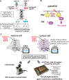
Fig. 1.. Probing dynamic signal transduction and filtering in cancer cells.
(A) Environmental stimuli can induce different dynamic patterns of Erk activity, which are then interpreted by downstream transcriptional circuits to specify cell behavior. (B) OptoSOS is an optogenetic method for Ras activation that enables probing of how cells filter and respond to dynamic Ras inputs. The light-inducible PhyB-PIF heterodimer drives membrane recruitment of the Ras-activating SOS2 catalytic domain, which activates Ras at the membrane. Red light (650 nm) induces PhyB-PIF dimerization, whereas far-red light (750 nm) dissociates the dimer. (C) We tested the hypothesis that some cancer cells may inappropriately filter dynamic Ras-Erk signals. We examined how dynamic optogenetic inputs were interpreted by normal or cancer cells through a combination of live-cell microscopy, high-throughput optogenetic stimulation (fig. S1), and immunofluorescence.
Fig. 2.
Fig. 2.. B-Raf mutant H1395 cells have an impaired transmission of pulsatile Ras signals.
(A) H1395 cells (bottom) showed extended kinetics of activation and inactivation in response to defined Ras input pulses. By contrast, NIH 3T3 cells (top) and the other cells in our cell line panel exhibited rapid kinetics (fig. S2B). Traces represent quantitation from live-cell imaging of nuclear BFP-Erk2 reporter accumulation. Traces were normalized between 0 and 1 and represent the mean ± 1 SD of 15 and 14 cells for 3T3 and H1395 cells, respectively. (B) Inactivation kinetics for H1395 and NIH 3T3 cells were confirmed through Western blot (blots are available in fig. S3A). Western blot quantification of ppErk is shown and fitted to single exponential decay.The dashed blue line depicts basal amount of ppErk from unstimulated cells. (C) Loss of fidelity in dynamic signal transduction in H1395 cells was observed through live-cell microscopy. 16-HBE (normal) and H1395 (cancer) cells were subjected to various dynamic patterns of input signal. (Three input conditions are shown. All six input conditions are shown in fig. S3B). As optoSOS input frequency increased, the H1395 cancer cells progressively lost their response to the gaps in the signal, whereas the normal cells did not. Traces represent the mean of five cells. Individual traces can be seen in fig. S3B. (D) Changes in the cell’s signal perception are analogous to cellular “blurred vision” for external stimuli. (Single-letter abbreviations for the amino acid residues are as follows: A, Ala; G, Gly; and V, Val. In the mutants, other amino acids were substituted at certain locations; for example, G469A indicates that glycine at position 469 is replaced by alanine.)
Fig. 3.
Fig. 3.. B-Raf P-loop mutations and drugs that perturb Raf dimerization both extend Ras-Erk pathway kinetics.
(A) OptoSOS and optoBRaf coupled with MEK inhibition (U0126) and mutant-B-Raf inhibition (PLX-8394) were used to isolate B-Raf as a network node that can extend Erk kinetics. Plots show quantification of Western blot data (blots are available in fig. S4, A, B, and C). Normal wild-type decay is indicated with the gray dashed line; basal signaling level (no opto-stimulation) is indicated by the purple dotted line. (B) Treatment of NIH 3T3s with paradoxically activating B-Raf inhibitors vemurafenib and SB590885 also extended ppErk decay kinetics. Datapoints show means ± 95% confidence interval (CI) of mean single-cell ppErk immunofluoresecnce from three replicates. (C) Our data support a model in which P-loop B-Raf mutations or paradoxically activating drugs can both enhance the Ras-induced dimerization potential of B-Raf and C-Raf, thus altering the kinetic properties of pathway activation and inactivation.
Fig. 4.
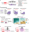
Fig. 4.. Perturbation of Ras-Erk signaling dynamics can alter how cells make proliferative decisions.
(A) Transcriptional decoding of dynamic signal inputs was examined in normal NIH 3T3 cells (fast pathway kinetics) or in cells treated with the kinetics-altering drug SB590885. Cells were stimulated with fixed-width signal pulses separated by various intervals. Expression of Erk targets and downstream cell-cycle entry were examined. (B) Altered Ras-Erk kinetics changed transcriptional output to dynamic Ras inputs. Immunofluorescence of cJun, EGR1, and Cyclin D1 expression time courses is depicted. Only expression in response to constant stimulus or a representative pulsed stimulus is shown.All input conditions tested are provided in fig. S11. Illumination was achieved with the optoPlate, and protein expression was assessed through single-cell immunofluorescence coupled with high-content imaging. Data points represent the median target fluorescence from 3000 to 4000 cells for each condition. (C) Extended Ras-Erk kinetics sensitized cells to proliferate under nonproliferative conditions. We used 384-well optoPlate illumination to examine proliferation of cells in response to a systematic scan of dynamic inputs. Normal and drug-treated cells were exposed to all combinations of six optoSOS pulse lengths (ON interval) and separated by seven pulse interval lengths (OFF interval) over 19 hours. Cells were then incubated with Edu for 30 min, fixed, stained, imaged, and analyzed. The percentage of cells incorporating Edu was plotted as an interpolated heatmap. Further analysis is available in figs. S12 and S13. The values used to generate the map represent means of biological quadruplicates. (D) Our data support the model that altering dynamic signal filtering properties can reshape the input-response map and may drive improper cellular behavior, such as hyperproliferation.

Comment in

  • From oncogenic mutation to dynamic code.
    Kolch W, Kiel C. Kolch W, et al. Science. 2018 Aug 31;361(6405):844-845. doi: 10.1126/science.aau8059. Science. 2018. PMID: 30166473 No abstract available.
  • Lighting Up Cancer Dynamics.
    Ravindran PT, Wilson MZ. Ravindran PT, et al. Trends Cancer. 2018 Oct;4(10):657-659. doi: 10.1016/j.trecan.2018.06.001. Epub 2018 Sep 25. Trends Cancer. 2018. PMID: 30292348

References

    1. Shaul YD, Seger R, The MEK/ERK cascade: From signaling specificity to diverse functions. Biochim. Biophys. Acta 1773, 1213–1226 (2007). - PubMed
    1. Albeck JG, Mills GB, Brugge JS, Frequency-modulated pulses of ERK activity transmit quantitative proliferation signals. Mol. Cell 49, 249–261 (2013). doi: 10.1016/j.molcel.2012.11.002 - DOI - PMC - PubMed
    1. Aoki K et al., Stochastic ERK activation induced by noise and cell-to-cell propagation regulates cell density-dependent proliferation. Mol. Cell 52, 529–540 (2013). doi: 10.1016/j.molcel.2013.09.015 - DOI - PubMed
    1. Hiratsuka T et al., Intercellular propagation of extracellular signal-regulated kinase activation revealed by in vivo imaging of mouse skin. eLife 4, e05178 (2015). doi: 10.7554/eLife.05178 - DOI - PMC - PubMed
    1. Marshall CJ, Specificity of receptor tyrosine kinase signaling: Transient versus sustained extracellular signal-regulated kinase activation. Cell 80, 179–185 (1995). doi: 10.1016/0092-8674(95)90401-8 - DOI - PubMed

Publication types

MeSH terms

Substances