

Visualizing and discovering cellular structures with super-resolution microscopy
- PMID: 30166485
- PMCID: PMC6535400
- DOI: 10.1126/science.aau1044
Visualizing and discovering cellular structures with super-resolution microscopy
Abstract
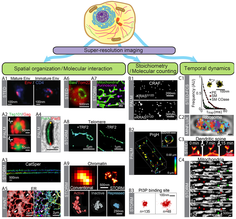
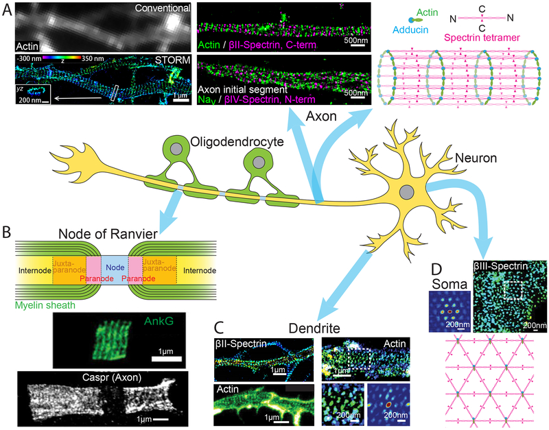
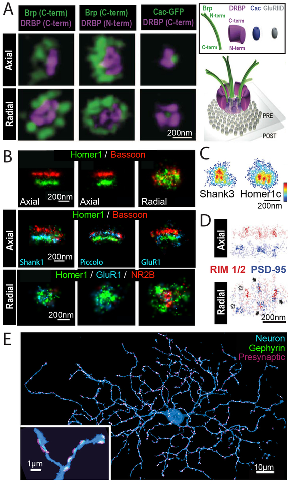
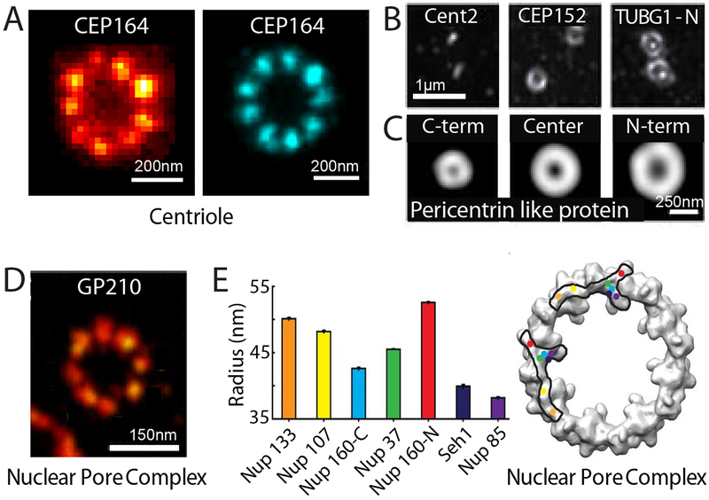
Super-resolution microscopy has overcome a long-held resolution barrier-the diffraction limit-in light microscopy and enabled visualization of previously invisible molecular details in biological systems. Since their conception, super-resolution imaging methods have continually evolved and can now be used to image cellular structures in three dimensions, multiple colors, and living systems with nanometer-scale resolution. These methods have been applied to answer questions involving the organization, interaction, stoichiometry, and dynamics of individual molecular building blocks and their integration into functional machineries in cells and tissues. In this Review, we provide an overview of super-resolution methods, their state-of-the-art capabilities, and their constantly expanding applications to biology, with a focus on the latter. We will also describe the current technical challenges and future advances anticipated in super-resolution imaging.
Copyright © 2018, American Association for the Advancement of Science.
Conflict of interest statement
Figures
References
-
- Hell SW, Wichmann J, Breaking the diffraction resolution limit by stimulated emission: stimulated-emission-depletion fluorescence microscopy. Optics letters 19, 780–782 (1994). - PubMed
-
- Klar TA, Hell SW, Subdiffraction resolution in far-field fluorescence microscopy. Optics letters 24, 954–956 (1999). - PubMed
-
- Eggeling C, Willig KI, Sahl SJ, Hell SW, Lens-based fluorescence nanoscopy. Quarterly reviews of biophysics 48, 178–243 (2015). - PubMed
-
- Heintzmann R, Gustafsson MGL, Subdiffraction resolution in continuous samples. Nature Photonics 3, 362–364 (2009).
Publication types
MeSH terms
Grants and funding
LinkOut - more resources
Full Text Sources
Other Literature Sources
