. 2018 Aug 30;9(1):3539.
doi: 10.1038/s41467-018-05916-9.

Luciferase-induced photoreductive uncaging of small-molecule effectors

Affiliations

Luciferase-induced photoreductive uncaging of small-molecule effectors

Eric Lindberg et al. Nat Commun. .

Abstract

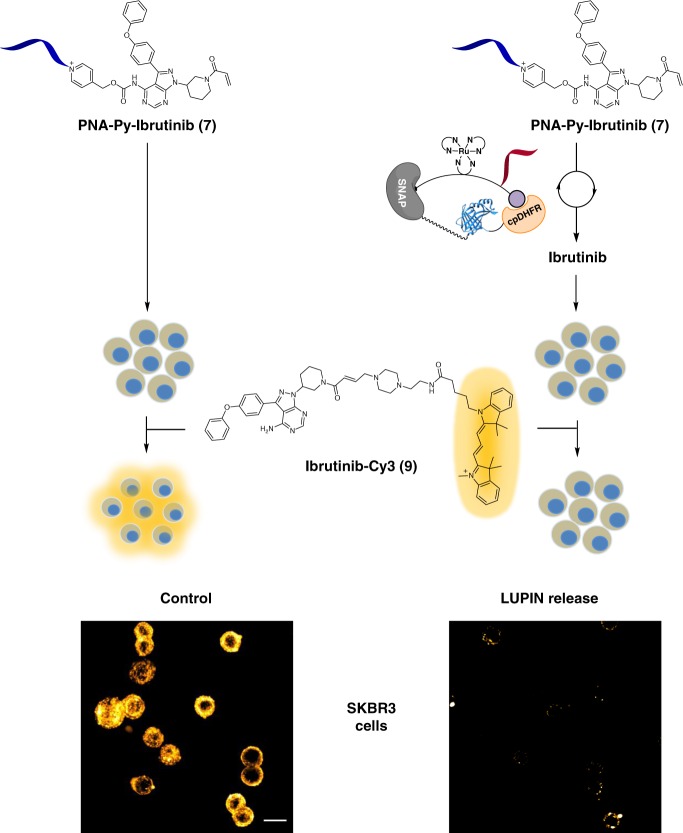
Bioluminescence resonance energy transfer (BRET) is extensively used to study dynamic systems and has been utilized in sensors for studying protein proximity, metabolites, and drug concentrations. Herein, we demonstrate that BRET can activate a ruthenium-based photocatalyst which performs bioorthogonal reactions. BRET from luciferase to the ruthenium photocatalyst is used to uncage effector molecules with up to 64 turnovers of the catalyst, achieving concentrations >0.6 μM effector with 10 nM luciferase construct. Using a BRET sensor, we further demonstrate that the catalysis can be modulated in response to an analyte, analogous to allosterically controlled enzymes. The BRET-induced reaction is used to uncage small-molecule drugs (ibrutinib and duocarmycin) at biologically effective concentrations in cellulo.


Conflict of interest statement

The authors declare no competing interests.

Figures

Fig. 1
Fig. 1
Concept and design of LUPIN. a The emission spectrum of NLuc (blue) overlaps very well with the absorption spectrum of the ruthenium photocatalyst (red), suggesting that efficient BRET should be possible. b The fusion protein SNAP-Pro30-NanoLuc (NLuc)-cpDHFR is linked via self-labeling SNAP to the synthetic linker containing the ruthenium photocatalyst and methotrexate (purple ball, DHFR ligand), positioning the ruthenium in close proximity to the NLuc. Free methotrexate (green ball) can push the sensor into the open conformation, thus turning off the BRET to the photocatalyst. c By installing a PNA next to the ruthenium catalyst, complementary pyridinium substrates can bind and undergo photoreductive cleavage by the ruthenium, unmasking the effector molecule
Fig. 2
Fig. 2
Characterization of LUPIN. a Bioluminescence resonance energy transfer from Nluc to ruthenium is observed when SNAP-NLuc-DHFR was labeled with BG-(Ru)(PNA)-MTX (1). Emergence of a new band at 610 nm indicates BRET from Nluc to ruthenium (red line); in the presence of methotrexate, more of the sensor shifts into the open conformation, decreasing BRET efficiency (black line). b Fluorescence enhancement due to rhodamine uncaging by the LUPIN system is observed. In the presence of methotrexate, the reaction is partially inhibited due to lower BRET efficiency. In the absence of nucleic acid template, or in the absence of a covalent link between the synthetic linker and the protein construct, the reaction is marginal. Reaction conditions: SNAP-NLuc-DHFR labeled with BG-(Ru)(PNA)-MTX (1) (50 nM), PNA-PyRho (2) or PrPyRho (3) (0.5 µM), sodium ascorbate (10 mM), furimazine (100 µM), and methotrexate (100 µM). BG benzyl guanine. c Varying the concentration of the labeled SNAP-NLuc-DHFR protein (50, 10, and 2 nM) has a significant impact on luminescence decay and the rate of rhodamine release
Fig. 3
Fig. 3
PNA-templated photoreductive release of effector molecules by ruthenium photocatalysis. Ru-PNA-5mer (5) (10 µM), PNA-Py-Drug (68) (100 µM), sodium ascorbate (10 mM) in PBS 1X(pH 7.4). Irradiation with 455 nm light before and after 2 min was followed by LC-MS quantification
Fig. 4
Fig. 4
LUPIN release of ibrutinib. Ibrutinib release by LUPIN was demonstrated by competition with ibrutinib-Cy3 (9) derivative in SKBR3 cells overexpressing ErbB2. SKBR3 cells were treated with either the prodrug alone (7) or prodrug (7) + LUPIN for 30 min; then the cells were treated with ibrutinib-Cy3 (9). The lower fluorescence observed for the LUPIN-uncaged product population indicates that the ErbB2 was saturated with the uncaged prodrug. Conditions for LUPIN release: SNAP-NLuc-DHFR labeled with BG-(Ru)(PNA)-MTX (1) (10 nM), PNA-PyIbr (7) (5 µM), sodium ascorbate (10 mM), furimazine (100 µM), and Ibr-Cy3 (9) (50 nM). Scale bar: 20 µm
Fig. 5
Fig. 5
LUPIN release of cytotoxic duocarmycin analogue in MCF-7 cell culture. a Schematic representation of Duo-OMe (4) release by LUPIN with furimazine to generate DNA alkylating agent. b Quantification of cellular growth (MCF-7) by nuclei count. Bar graph of nuclei count across different furimazine and prodrug (PNA-Py2Duo 12) concentrations (left panel). Representative images of cells (right panel) across different prodrug (12) and furimazine concentrations imaged by fluorescence (Hoechst) and bright field (BF). The red square indicates the zoomed-in area below it. The bar graph is the average of three independent experiments ran in triplicates. Error bars show ±1 standard deviation from the mean. Statistics were calculated using a two-tailed t-test with unequal variances (Welch’s unpaired t-test). *p < 0.05, **p < 0.01, ***p < 0.001. Scale bar: 200 µM; zoom-in scale bar: 50 µM

References

    1. Aper SJA, Dierickx P, Merkx M. Dual readout BRET/FRET sensors for measuring intracellular zinc. ACS Chem. Biol. 2016;11:2854–2864. doi: 10.1021/acschembio.6b00453. - DOI - PMC - PubMed
    1. Dragulescu-Andrasi A, Chan CT, De A, Massoud TF, Gambhir SS. Bioluminescence resonance energy transfer (BRET) imaging of protein-protein interactions within deep tissues of living subjects. Proc. Nat. Acad. Sci. 2011;108:12060–12065. doi: 10.1073/pnas.1100923108. - DOI - PMC - PubMed
    1. Goyet E, Bouquier N, Ollendorff V, Perroy J. Fast and high resolution single-cell BRET imaging. Sci. Rep. 2016;6:28231. doi: 10.1038/srep28231. - DOI - PMC - PubMed
    1. Machleidt T, et al. NanoBRET--A novel BRET platform for the analysis of protein-protein interactions. ACS Chem. Biol. 2015;10:1797–1804. doi: 10.1021/acschembio.5b00143. - DOI - PubMed
    1. Schaub FX, et al. Fluorophore-NanoLuc BRET reporters enable sensitive in vivo optical imaging and flow cytometry for monitoring tumorigenesis. Cancer Res. 2015;75:5023–5033. doi: 10.1158/0008-5472.CAN-14-3538. - DOI - PMC - PubMed

Publication types

MeSH terms