

MITOCHONDRIAL DYNAMICS AND METABOLIC REGULATION IN CARDIAC AND SKELETAL MUSCLE
- PMID: 30166722
- PMCID: PMC6116613
MITOCHONDRIAL DYNAMICS AND METABOLIC REGULATION IN CARDIAC AND SKELETAL MUSCLE
Abstract
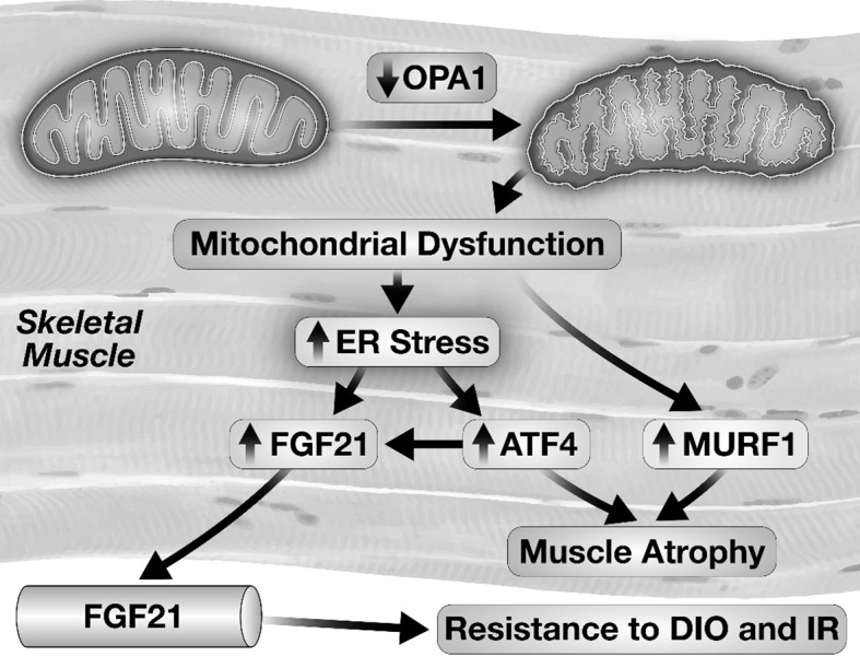
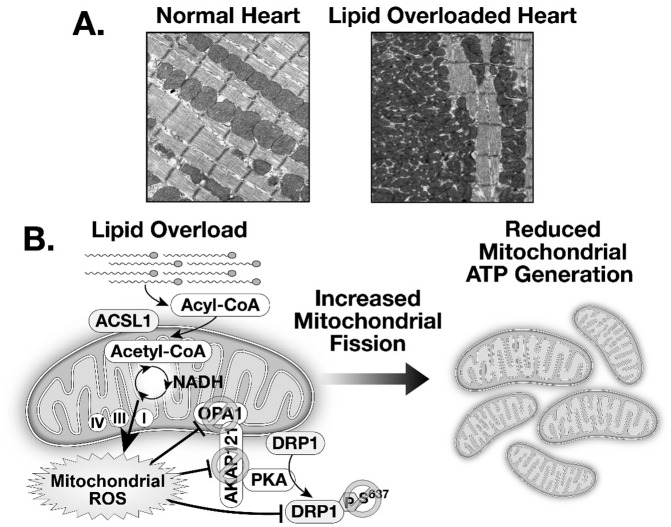
Mitochondria are the metabolic powerhouses of cells. In addition to generating adenosine triphosphate (ATP), they play important roles in cell survival pathways such as apoptosis and necrosis. Mitochondrial size and shape are dynamically regulated by a process known as mitochondrial dynamics. The significance of this process in metabolically active cells such as skeletal and cardiac muscle are only now beginning to be elucidated. In cardiac muscle, mitochondrial dynamics plays an important role in mitochondrial quality control and defects in regulatory pathways that govern these processes and leads to heart failure. In response to nutrient excess such as lipid overload, as occurs in diabetes, mitochondrial shape and morphology are altered by effects of nutrient stress on mitochondrial dynamics signaling pathways, which have important implications for understanding mitochondrial dysfunction in diabetic cardiomyopathy. Moreover, crosstalk between mitochondria and other organelles such as the endoplasmic reticulum can regulate generation of hormones such as fibroblast growth factor 21, with potent anti-diabetic and anti-obesity effects.
Conflict of interest statement
Potential Conflicts of Interest: None disclosed.
Figures
References
-
- Liesa M, Palacin M. Zorzano A. Mitochondrial dynamics in mammalian health and disease. Physiol Rev. 2009;89:799–845. - PubMed
-
- Pernas L. Scorrano L. Mito-morphosis: mitochondrial fusion, fission, and cristae remodeling as key mediators of cellular function. Annu Rev Physiol. 2016;78:505–31. - PubMed
-
- Anton F, Dittmar G, Langer T. Escobar-Henriques M. Two deubiquitylases act on mitofusin and regulate mitochondrial fusion along independent pathways. Mol Cell. 2013;49:487–98. - PubMed
-
- Wai T. Langer T. Mitochondrial dynamics and metabolic regulation. Trends Endocrinol Metab. 2016;27:105–17. - PubMed
MeSH terms
Substances
LinkOut - more resources
Full Text Sources
Other Literature Sources
Medical