

From electron crystallography of 2D crystals to MicroED of 3D crystals
- PMID: 30166936
- PMCID: PMC6112780
- DOI: 10.1016/j.cocis.2018.01.010
From electron crystallography of 2D crystals to MicroED of 3D crystals
Abstract
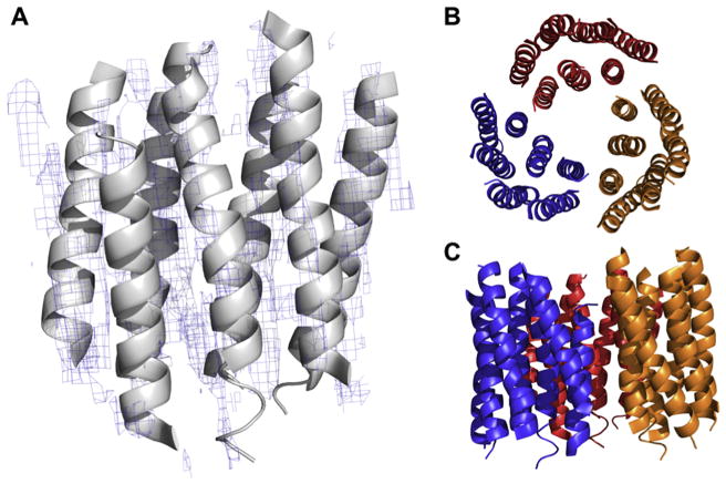
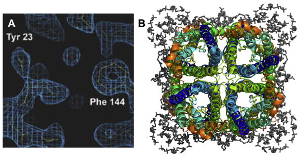
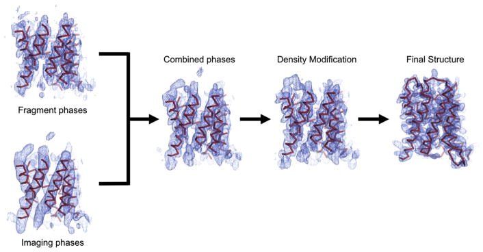
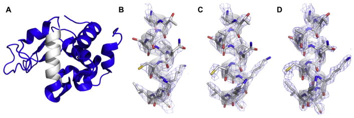
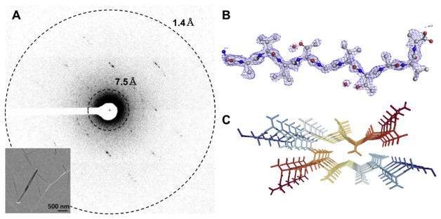
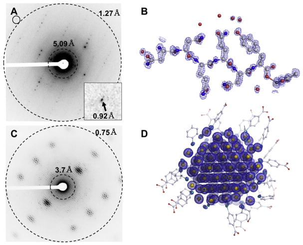
Electron crystallography is widespread in material science applications, but for biological samples its use has been restricted to a handful of examples where two-dimensional (2D) crystals or helical samples were studied either by electron diffraction and/or imaging. Electron crystallography in cryoEM, was developed in the mid-1970s and used to solve the structure of several membrane proteins and some soluble proteins. In 2013, a new method for cryoEM was unveiled and named Micro-crystal Electron Diffraction, or MicroED, which is essentially three-dimensional (3D) electron crystallography of microscopic crystals. This method uses truly 3D crystals, that are about a billion times smaller than those typically used for X-ray crystallography, for electron diffraction studies. There are several important differences and some similarities between electron crystallography of 2D crystals and MicroED. In this review, we describe the development of these techniques, their similarities and differences, and offer our opinion of future directions in both fields.
Keywords: Electron diffraction; MicroED; cryoEM (electorn cryo-microscopy); electron crystallography.
Figures
References
-
- Doerr A. Cryo-electron tomography. Nat Methods. 2017;14:34. doi: 10.1038/nmeth.4115. - DOI
Grants and funding
LinkOut - more resources
Full Text Sources
Other Literature Sources