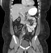
Gallbladder agenesis mimicking cholelithiasis in an adult
- PMID: 30167026
- PMCID: PMC6114116
- DOI: 10.1016/j.radcr.2018.03.002
Gallbladder agenesis mimicking cholelithiasis in an adult
Abstract
We present the case of a 24-year-old woman with morbid obesity who came to the emergency department with right upper quadrant abdominal pain associated with nausea and vomiting. Her workup included a right upper quadrant ultrasound suggestive of a small gallbladder with cholelithiasis without sonographic evidence of acute cholecystitis. She underwent attempted laparoscopic cholecystectomy with no identifiable gallbladder during surgery. Postsurgical cross-sectional imaging confirmed gallbladder agenesis. This case provides an example of a rare but convincing clinical and radiologic mimic of cholelithiasis. In certain cases of biliary colic and imaging revealing a small gallbladder, a magnetic resonance cholangiopancreatography may be warranted to evaluate gallbladder agenesis and avoid unnecessary surgery.
Keywords: Cholelithiasis; Gallbladder agenesis; Magnetic resonance cholangiopancreatography; Ultrasound.
Figures


References
-
- Bennion R.S., Thompson J.E., Jr, Tompkins R.K. Agenesis of the gallbladder without extrahepatic biliary atresia. Arch Surg. 1988;123(10):1257–1260. - PubMed
-
- Senecail B., Texier F., Kergastel L., Paton-Philippe L. Anatomic variability and congenital anomalies of the gallbladder and biliary tract: ultrasonographic study of 1823 patients. Morphologie. 2000;84:35–39. - PubMed
-
- Toouli J., Geenen J.E., Hogan W.J., Dodds W.J., Arndorfer R.C. Sphincter of Oddi motor activity: a comparison between patients with common bile duct stones and controls. Gastroenterology. 1982;82(1):111–117. - PubMed
Publication types
LinkOut - more resources
Full Text Sources
Other Literature Sources

