. 2018 Aug 3;9(60):31753-31770.
doi: 10.18632/oncotarget.25878.

A novel fatty acid-binding protein 5-estrogen-related receptor α signaling pathway promotes cell growth and energy metabolism in prostate cancer cells

Affiliations

A novel fatty acid-binding protein 5-estrogen-related receptor α signaling pathway promotes cell growth and energy metabolism in prostate cancer cells

Shogo Senga et al. Oncotarget. .

Abstract

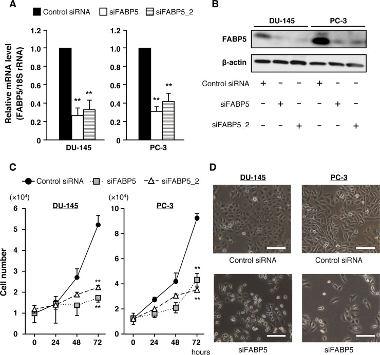
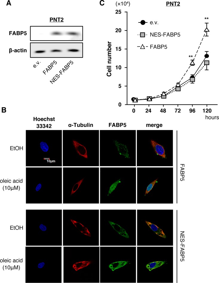
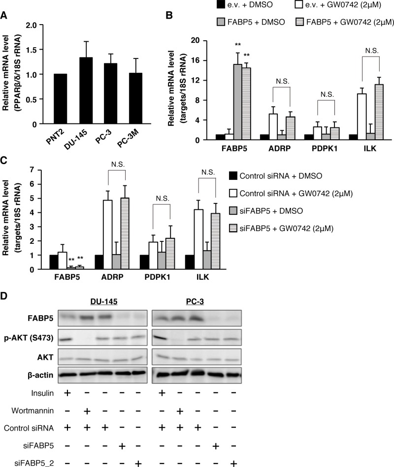
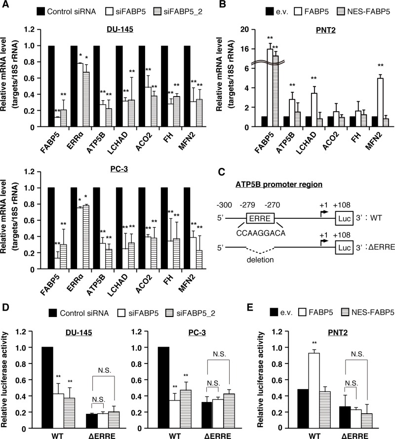
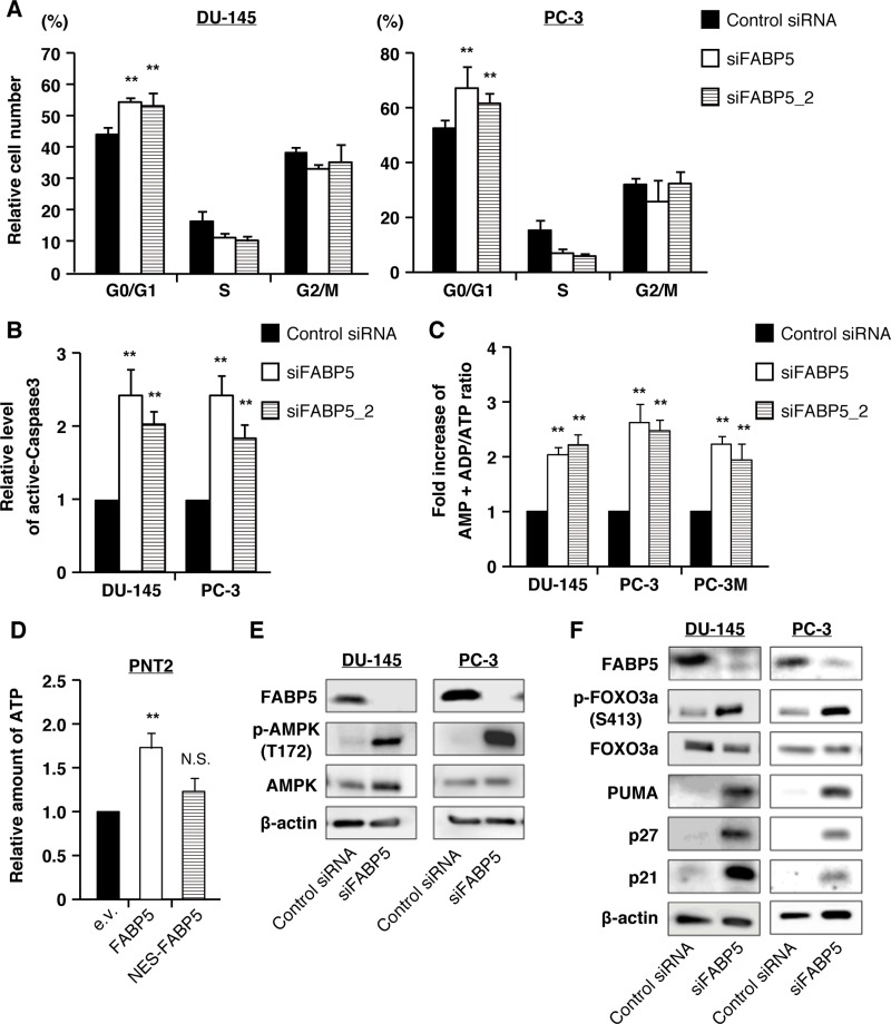
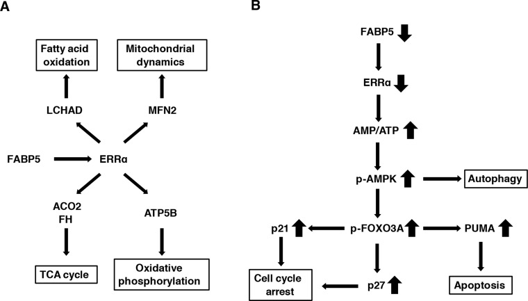
Epidermal or cutaneous fatty acid-binding protein is an intracellular lipid-binding protein, also known as FABP5, and its expression level is closely related to cancer cell proliferation and metastatic activities in various types of carcinoma. However, the molecular mechanisms of FABP5 in cancer cell proliferation and its other functions have remained unclear. In the present study, we have clearly revealed that FABP5 activated expression of metabolic genes (ATP5B, LCHAD, ACO2, FH and MFN2) via a novel signaling pathway in an ERRα (estrogen-related receptor α)-dependent manner in prostate cancer cell lines. To clarify the novel function of FABP5, we examined the activation mechanisms of the ERRα target genes via FABP5. A direct protein-protein interaction between FABP5 and ERRα was demonstrated by immunoprecipitation and GST pull-down assays. We have clearly revealed that FABP5 interacted directly with transcriptional complex containing ERRα and its co-activator PGC-1β to increase expression of the ERRα target genes. In addition, we have shown that FABP5 knockdown induced high energy stress leading to induction of apoptosis and cell cycle arrest via AMPK-FOXO3A signaling pathway in prostate cancer cells, suggesting that FABP5 plays an important role in cellular energy status directing metabolic adaptation to support cellular proliferation and survival.

Keywords: ERRα; FABP5; PGC-1β; energy metabolism; prostate cancer.


Conflict of interest statement

CONFLICTS OF INTEREST The authors declare no conflicts of interest.

Figures

Figure 1
Figure 1. FABP5 knockdown significantly suppresses cell growth in PCa cells
(A) FABP5 mRNA levels in PCa cells transfected with control siRNA (20 nM) or siFABP5 (20 nM) determined by qPCR. Results are means ± S.D. for three independent experiments. (B) Western blot analysis of FABP5 in PCa cells transfected with control siRNA (20 nM) or siFABP5 (20 nM). β-actin served as an internal loading control. Results shown are representative of three independent experiments. (C) Cell proliferation of control siRNA (20 nM) or siFABP5 (20 nM) transfected PCa cells. Cells were counted at the indicated times. Results are means ± S.D. for three independent experiments. (D) Representative images of cells transfected with control siRNA (upper panel) or siFABP5 (lower panel) 72 h after transfection. Scale bar, 200 μm. **P < 0.01, one-way ANOVA followed by Dunnett's test (A), two-way ANOVA followed by Tukey's test (C).
Figure 2
Figure 2. Nuclear localization of FABP5 promotes cell proliferation
(A) Western blot analysis of FABP5 and NES-FABP5 in PNT2 cells transfected with pCI-neo/FABP5, NES-FABP5 or pCI-neo (e.v., empty vector). Results shown are representative of three independent experiments. (B) Localization of FABP5 and NES-FABP5 in FABP5 or NES-FABP5 overexpressing PNT2 cells. Transfected cells were treated with ethanol or oleic acid (10 μM) for 30 min. (C) Cell growth of FABP5- and NES-FABP5-transfected PNT2 cells. Cells were counted at the indicated times. Results are means ± S.D. for three independent experiments. **P < 0.01, two-way ANOVA followed by Tukey's test.
Figure 3
Figure 3. PPARβ/δ signaling might not be involved in FABP5-mediated growth promotion of PCa cells
(A) PPARβ/δ mRNA expression levels in PNT2, DU-145, PC-3 and PC-3M. Relative mRNA levels were measured by qPCR. Results are means ± S.D. for three independent experiments. (B) PNT2 cells were transfected with pCI-neo or pCI-neo/FABP5. At 48 h after transfection, transfected cells were treated with DMSO or GW0742 (2 μM) for 24 h. mRNA levels of FABP5, PDPK1, ADRP and ILK were measured by qPCR and normalized to those of 18S rRNA. Results are means ± S.D. for three independent experiments. (C) PC-3 cells were transfected with control siRNA (20 nM) or siFABP5 (20 nM). At 48 h after transfection, cells were treated with DMSO or GW0742 (2 μM) for 24 h. FABP5, PDPK1, ADRP and ILK mRNA levels were measured by qPCR and normalized to those of 18S rRNA. Results are means ± S.D. for three independent experiments. (D) Phosphorylation of Akt (S473) in siFABP5 transfected DU-145 and PC-3 was detected by western blot analysis. We used insulin (as positive control of AKT phosphorylation, 1 μg/ml, 15 min) and wortmannin (inhibitor of phosphoinositide 3-kinases, as negative control of AKT phosphorylation, 0.5 μM, 30 min). Results shown are representative of three independent experiments. **P < 0.01, one-way ANOVA followed by Tukey's test (B and C), N.S. = Not significant.
Figure 4
Figure 4. FABP5 promotes cell growth via ERRα signaling pathway in PCa cells
(A) PCa cells were transfected with control siRNA (20 nM) or siFABP5 (20 nM) and mRNA levels of various factors were determined by qPCR. Results are means ± S.D. for three independent experiments. (B) PNT2 cells were transfected with pCI-neo (e.v.), pCI-neo/FABP5 (FABP5) or pCI-neo/NES-FABP5 (NES-FABP5) and mRNA levels of various factors were measured by qPCR. Results are means ± S.D. for three independent experiments. (C) Schematic of the ATP5B promoter region and luciferase constructs with the wild-type (WT) sequence or mutant deleted for the ERRE (ΔERRE). (D) The luciferase reporter vector driven by the ATP5B promoter (WT) or ΔERRE mutant was co-transfected with hRluc/TK into PC-3 and DU-145 cells, and treated with control siRNA (20 nM) or siFABP5 (20 nM). Luciferase activities were measured and normalized against Renilla activities. Results are means ± S.D. for three independent experiments. (E) The WT or ΔERRE mutant reporter vector was co-transfected with hRluc/TK into PNT2 cells transfected with pCI-neo (e.v.), pCI-neo/FABP5 or NES-FABP5. Luciferase activities were measured and normalized against Renilla activities. Results are means ± S.D. for three independent experiments. *P < 0.05, **P < 0.01, one-way ANOVA followed by Dunnett's test (A, B, D and E), N.S. = Not significant.
Figure 5
Figure 5. FABP5 activates ERRα target genes mediated by direct interaction with ERRα in the nucleus
(A) Detection of ERK8 expression level in control siRNA (20 nM) or siFABP5 (20 nM) transfected PCa cells by western blot analysis. The arrow indicates ERK8 band. Results shown are representative of three independent experiments. (B) Western blot analysis of ERRα in nuclear and cytoplasmic fractions from control siRNA (20 nM) or siFABP5 (20 nM) transfected PC-3 cells. TBP served as a nuclear marker and tubulin served as cytoplasmic marker. (C, D) GST pull-down assays. ERRα contains an activation function 1 (AF-1, 1~75aa), a central zinc finger DNA binding domain (DBD, 75~151aa), a hinge region (151~225aa), a ligand-binding domain (LBD, 225~423aa), and an activation function 2 domain (AF-2, 398~423aa). We prepared two deletion mutants of ERRα (Δ300aa and Δ398aa). GST, GST-ERRα, GST-ERRα Δ300aa or GST-ERRα Δ398aa was incubated with PC-3 whole cell lysates and glutathione sepharose overnight at 4°C. GST, FABP5 and PGC-1β levels were detected by western blot analysis. Results shown are representative of three independent experiments. (E) Immunoprecipitation was performed with FLAG antibody (MBL) on lysates from pCI-neo (e.v.) or pCI-neo/3×FLAG-FABP5-transfected 293T cells. Input (10%) and IP samples were analyzed by western blot analysis. Results shown are representative of three independent experiments. (F) ChIP–qPCR analysis to evaluate binding of the FABP5-ERRα complex to the ATP5B promoter. 293T cells were transfected with pCI-neo (e.v.), FLAG-FABP5, ERRα and pGC-1β as indicated. ChIP was performed using anti-DDDDK-tag antibody, and the ERRα-binding site in ATP5B (−300 to −213) promoter region was amplified using ChIP-qPCR primers to calculate % input. Primer sequences are described in the SI Materials and Methods. Results are mean ± S.D. for three independent experiments. **P < 0.01, one-way ANOVA followed by Tukey's test.
Figure 6
Figure 6. FABP5 knockdown induces starvation stress and activation of the AMPK-FOXO3A signaling pathway in PCa cell lines
(A) Cell cycle analysis of control siRNA (20 nM) or siFABP5 (20 nM) transfected PCa cells by flow cytometry. Percentages of cells at each phase are indicated. (B) Apoptosis was measured by cleaved caspase 3 (active form) level by flow cytometry. (C) Amounts of AMP+ADP and ATP were measured by the microplate reader using firefly luciferase in control siRNA (20 nM) or siFABP5 (20 nM) transfected PCa cells. Data are presented as the means ± SD from four independent experiments. (D) Relative amount of ATP was measured using firefly luciferase in pCI-neo (e.v.), pCI-neo/FABP5 or pCI-neo/NES-FABP5-transfected PNT2 cells. Results are mean ± S.D. for twenty independent experiments. (E) Western blot analysis of AMPK and phosphorylated AMPK in prostate cancer cells transfected with control siRNA (20 nM) or siFABP5 (20 nM). Results shown are representative of three independent experiments. (F) Western blot analysis of FOXO3A and proteins encoded by its target genes in PCa cells transfected with control siRNA (20 nM) or siFABP5 (20 nM). Results shown are representative of three independent experiments. **P < 0.01, two-way ANOVA followed by Tukey's test (A), one-way ANOVA followed by Dunnett's test (B, C and D), N.S. = Not significant.
Figure 7
Figure 7. Schematic model for the FABP5-mediated transcriptional controlling network of mitochondrial functions in PCa cells
(A) FABP5-ERRα crosstalk is required for mitochondrial functions and cell proliferation in PCa cells. FABP5 activated mRNA expression of more than one metabolic gene via ERRα signaling in PCa cells. (B) FABP5 knockdown induced high energy stress (increase of AMP/ATP ratio) in PCa cells. AMPK was activated by increased AMP/ATP ratio. Moreover, autophagy, apoptosis and cell cycle arrest were induced in PCa cells transfected with siRNA against FABP5 via the AMPK-FOXO3A signaling pathway. Thus, we considered that metabolic genes regulated by FABP5-ERRα played an essential role for energy homeostasis and viability in PCa cells. These results suggested that FABP5 may be a molecular target for the antitumorigenic treatments for PCa.

Similar articles

Cited by

References

    1. Siegel R, Ward E, Brawley O, Jemal A. Cancer statistics, 2011: the impact of eliminating socioeconomic and racial disparities on premature cancer deaths. CA Cancer J Clin. 2011;61:212–36. - PubMed
    1. Siegel RL, Miller KD, Jemal A. Cancer statistics, 2015. CA Cancer J Clin. 2015;65:5–29. - PubMed
    1. Kolonel LN, Nomura AM, Cooney RV. Dietary fat and prostate cancer: current status. J Natl Cancer Inst. 1999;91:414–28. - PubMed
    1. Suburu J, Chen YQ. Lipids and prostate cancer. Prostaglandins Other Lipid Mediat. 2012;98:1–10. - PMC - PubMed
    1. Swinnen JV, Roskams T, Joniau S, Van Poppel H, Oyen R, Baert L, Heyns W, Verhoeven G. Overexpression of fatty acid synthase is an early and common event in the development of prostate cancer. Int J Cancer. 2002;98:19–22. - PubMed