

Advanced Unilateral Fibrous Dysplasia of the Scapula: A Rare Clinical Entity and Surgical Challenge
- PMID: 30167424
- PMCID: PMC6114216
- DOI: 10.13107/jocr.2250-0685.1068
Advanced Unilateral Fibrous Dysplasia of the Scapula: A Rare Clinical Entity and Surgical Challenge
Abstract
Introduction: Fibrous dysplasia (FD) is an uncommon benign tumor of bone. Although FD can affect flat bones, it is rare for the scapula to be involved. In addition, little is known about the management of FD when it involves the scapula. We present possibly the first comprehensive case report of the management of advanced unilateral FD of the scapular region.
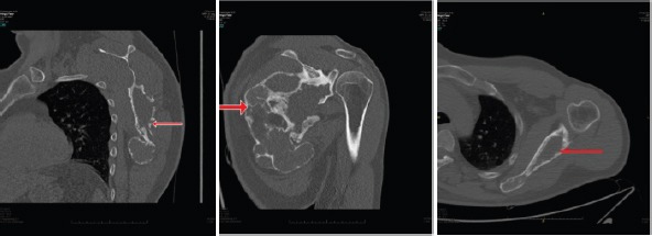
Case report: A 47-year-old male presented to us with pain and swelling over the left shoulder. The swelling was 11 cm × 15 cm × 8 cm and was hard and tender with rough texture. Radiograph showed large homogenous lesion with irregular but well-defined margins and a ground glass appearance. Magnetic resonance imaging scans showed well-defined borders with the expansion of the bone, with intact overlying cortices and endosteal scalloping. Biopsy confirmed the lesion to be FD. An innovative application of an existing surgical technique to minimize the impact of the residual deformity and dead space left after curettage of the scapula was done. The patient had good clinical and functional outcome at 6-month follow-up.
Conclusion: Surgical exercise in FD is purely on symptomatic basis. In our case, the swelling was causing most discomfort, and we curettaged and compressed the bony swelling which resulted in excellent outcome in this patient.
Keywords: Fibrous dysplasia; functional outcome; rare scapula lesion; unilateral fibrous dysplasia; unique surgical technique.
Conflict of interest statement
Conflict of Interest: Nil
Figures


Similar articles
-
A Rare Case of Fibrous Dysplasia Presenting With Facial Swelling and Craniofacial Deformity in a 13-Year-Old Girl.Cureus. 2024 Apr 29;16(4):e59327. doi: 10.7759/cureus.59327. eCollection 2024 Apr. Cureus. 2024. PMID: 38817487 Free PMC article.
-
Fibrous dysplasia of the anterior mandible: A rare case report.Tzu Chi Med J. 2018 Jul-Sep;30(3):185-187. doi: 10.4103/tcmj.tcmj_57_18. Tzu Chi Med J. 2018. PMID: 30069129 Free PMC article.
-
Pseudo-winging of Scapula due to Ventral Scapular Osteochondroma: A Case Report and Literature Review.J Orthop Case Rep. 2020 Aug-Sep;10(5):69-72. doi: 10.13107/jocr.2020.v10.i05.1844. J Orthop Case Rep. 2020. PMID: 33312984 Free PMC article.
-
Computed Tomography in Craniofacial Fibrous Dysplasia: A Case Series with Review of Literature and Classification Update.Open Dent J. 2017 Jun 30;11:384-403. doi: 10.2174/1874210601711010384. eCollection 2017. Open Dent J. 2017. PMID: 28839487 Free PMC article. Review.
-
Malignant transformation in monostotic fibrous dysplasia: clinical features, imaging features, outcomes in 10 patients, and review.Medicine (Baltimore). 2015 Jan;94(3):e369. doi: 10.1097/MD.0000000000000369. Medicine (Baltimore). 2015. PMID: 25621678 Free PMC article. Review.
Cited by
-
Shoulder arthroplasty in the setting of polyostotic fibrous dysplasia.JSES Rev Rep Tech. 2020 Nov 16;1(1):69-73. doi: 10.1016/j.xrrt.2020.100002. eCollection 2021 Feb. JSES Rev Rep Tech. 2020. PMID: 37588631 Free PMC article. No abstract available.
-
Intraosseous Pneumatocysts of the Scapula Mimicking Bone Tumors: A Report of Two Rare Cases Along with Elucidation of Their Etiology.Diseases. 2025 May 27;13(6):170. doi: 10.3390/diseases13060170. Diseases. 2025. PMID: 40558581 Free PMC article.
References
-
- Baig R, Eady JL. Unicameral (simple) bone cysts. South Med J. 2006;99:966–76. - PubMed
-
- DiCaprio MR, Enneking WF. Fibrous dysplasia. Pathophysiology, evaluation, and treatment. J Bone Joint Surg Am. 2005;87:1848–64. - PubMed
-
- Lichtenstein L. Polyostotic fibrous dysplasia. Arch Surg. 1938;36:874–98.
-
- Gass JD. Orbital and ocular involvement in fibrous dysplasia. South Med J. 1965;58:324–9. - PubMed
-
- Yabut SM, Jr, Kenan S, Sissons HA, Lewis MM. Malignant transformation of fibrous dysplasia. A case report and review of the literature. Clin Orthop Relat Res. 1988;228:281–9. - PubMed
Publication types
LinkOut - more resources
Full Text Sources