. 2018 Aug 29;4(8):eaau1291.
doi: 10.1126/sciadv.aau1291. eCollection 2018 Aug.

Electrophoretic drug delivery for seizure control

Affiliations

Electrophoretic drug delivery for seizure control

Christopher M Proctor et al. Sci Adv. .

Abstract

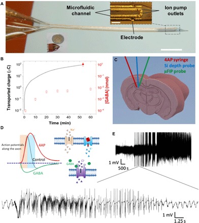
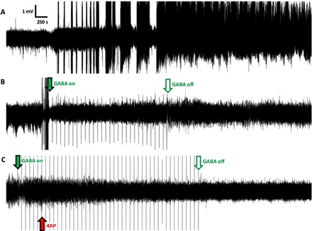
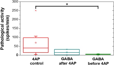
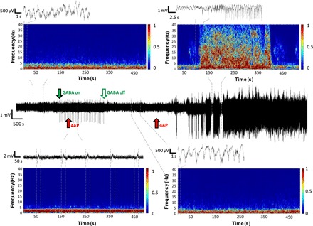
The persistence of intractable neurological disorders necessitates novel therapeutic solutions. We demonstrate the utility of direct in situ electrophoretic drug delivery to treat neurological disorders. We present a neural probe incorporating a microfluidic ion pump (μFIP) for on-demand drug delivery and electrodes for recording local neural activity. The μFIP works by electrophoretically pumping ions across an ion exchange membrane and thereby delivers only the drug of interest and not the solvent. This "dry" delivery enables precise drug release into the brain region with negligible local pressure increase. The therapeutic potential of the μFIP probe is tested in a rodent model of epilepsy. The μFIP probe can detect pathological activity and then intervene to stop seizures by delivering inhibitory neurotransmitters directly to the seizure source. We anticipate that further tailored engineering of the μFIP platform will enable additional applications in neural interfacing and the treatment of neurological disorders.


Figures

Fig. 1
Fig. 1. Overview of the μFIP probe.
(A) Implanted end of the device (inset scale bar, 100 μm; outside scale bar, 1 mm). (B) Net transported charge across the ion bridge when actively pumping GABA at 1 V (line, left axis), [GABA] passively diffused out of the device when no voltage was applied (open symbols, right axis), and [GABA] actively pumped out of the device at 1 V (closed symbols, right axis). (C) Schematic showing placement of syringe for 4AP injection, Si depth probe, and the μFIP probe in the hippocampus. (D) Conceptual illustration showing a proposed effect of 4AP on K+ channels and action potentials (31) along with the analogous effects of GABA. (E) Representative recording of intense SLEs following injection of 4AP at two different time scales.
Fig. 2
Fig. 2. Representative electrophysiology recordings from the hippocampus.
(A) Recording in the absence of the μFIP treatment with SLEs starting approximately 30 min after 4AP injection followed by status epilepticus. (B) Recording of case in which the μFIP treatment was initiated immediately following the first SLE, showing no further pathological events after the treatment starts. (C) Recording in which the μFIP treatment was initiated before 4AP injection, showing no pathological events. Red arrow indicates 4AP injection. Solid green arrows indicate start of the μFIP treatment, and open green arrows mark the end of the μFIP treatment. Sharp peaks at 100-s intervals following green arrow are artifacts from the μFIP treatment.
Fig. 3
Fig. 3. Frequency of pathological activity recorded for control experiments with only 4AP as well as for the case of delivering GABA after 4AP and the case of GABA before 4AP.
Fig. 4
Fig. 4. Representative recording from an anesthetized mouse given a 4AP injection during a period of GABA delivery (solid green arrow until open green arrow) followed by a second 4AP dose administered several minutes after the μFIP treatment was stopped.
Time/frequency plots for periods before (top left), during (bottom left), and after GABA delivery (bottom right) as well as during an SLE event (top right) are shown along with recording windows for shorter time scales. Dashed lines indicate the time periods covered by each time/frequency and recording plot.
Fig. 5
Fig. 5. Representative histological evaluation of implant traces.
(A) μFIP implant, (B) 4AP syringe, and (C) multichannel Si electrode in the hippocampus. The implants were labeled with DiI and shown as red traces. Astrocytes were labeled in green [glial fibrillary acidic protein (GFAP)], while all cellular nuclei were stained in blue [4′,6-diamidino-2-phenylindole (DAPI)]. Major regions of the hippocampus were labeled in white [cornu ammonis 1 (CA1), cornu ammonis 3 (CA3), and dentate gyrus (DG)]. Scale bar, 500 μm.

References

    1. Paz J. T., Paz J. T., Davidson T. J., Frechette E. S., Delord B., Parada I., Peng K., Deisseroth K., Huguenard J. R., Closed-loop optogenetic control of thalamus as a tool for interrupting seizures after cortical injury. Nat. Neurosci. 16, 64–70 (2013). - PMC - PubMed
    1. Armstrong C., Krook-Magnuson E., Oijala M., Soltesz I., Closed-loop optogenetic intervention in mice. Nat. Protoc. 8, 1475–1493 (2013). - PMC - PubMed
    1. Krook-Magnuson E., Armstrong C., Oijala M., Soltesz I., On-demand optogenetic control of spontaneous seizures in temporal lobe epilepsy. Nat. Commun. 4, 1376 (2013). - PMC - PubMed
    1. Bui A. D., Nguyen T. M., Limouse C., Kim H. K., Szabo G. G., Felong S., Maroso M., Soltesz I., Dentate gyrus mossy cells control spontaneous convulsive seizures and spatial memory. Science 359, 787–790 (2018). - PMC - PubMed
    1. Dong S., Rogan S. C., Roth B. L., Directed molecular evolution of DREADDs: A generic approach to creating next-generation RASSLs. Nat. Protoc. 5, 561–573 (2010). - PubMed

Publication types

MeSH terms