Review
. 2018 Oct 1;75(10):1273-1281.
doi: 10.1001/jamaneurol.2018.1073.

Advances in Understanding the Pathophysiology of Lacunar Stroke: A Review

Affiliations
Review

Advances in Understanding the Pathophysiology of Lacunar Stroke: A Review

Robert W Regenhardt et al. JAMA Neurol. .

Abstract

Importance: Stroke is the second leading cause of death in the world, and nearly one-third of ischemic strokes are lacunar strokes (LSs) or small subcortical infarcts. Although smaller in size, they create large problems, leaving many patients with intellectual and physical disabilities. Because there are limitations in understanding the underlying pathophysiology of LS, the development of novel therapies has been slow.

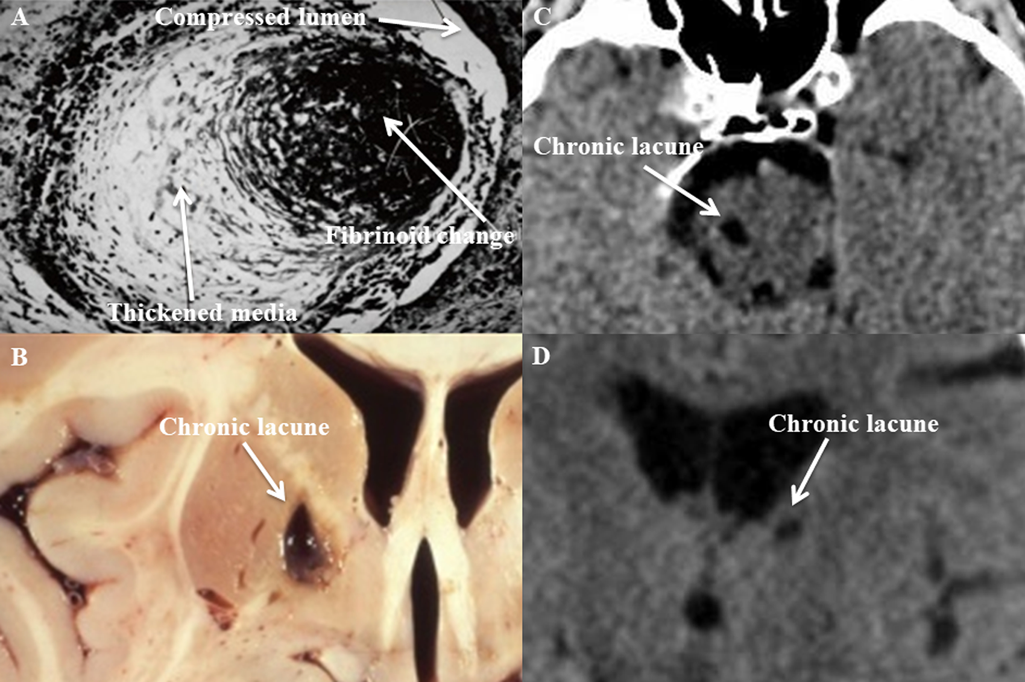
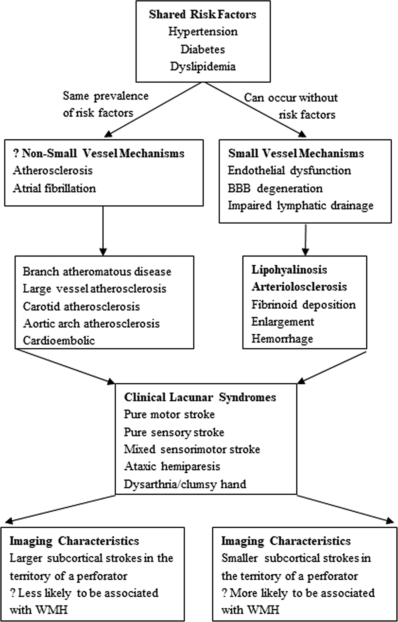
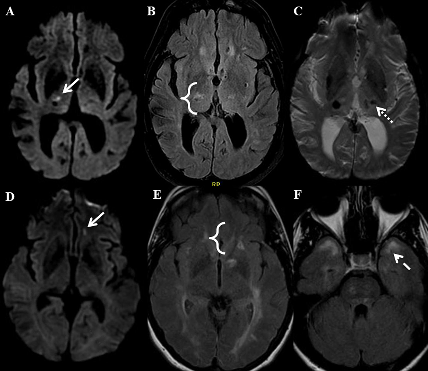
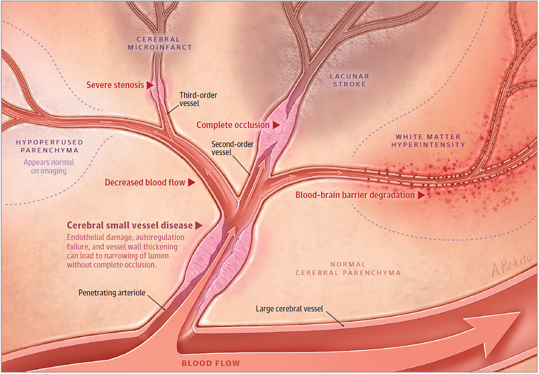
Observations: When the term lacune was described in the 1800s, its underlying pathophysiological basis was obscure. In the 1960s, C. Miller Fisher, MD, performed autopsy studies that showed that vessels supplying lacunes displayed segmental arteriolar disorganization, characterized by vessel enlargement, hemorrhage, and fibrinoid deposition. For these pathologic changes, he coined the term lipohyalinosis. Since that time, few attempts have been made to reconcile this pathologic description with modern mechanisms of cerebral small vessel disease (CSVD). During the past 6 years, progress has been made in understanding the clinical mechanisms, imaging characteristics, and genetic basis of LS.

Conclusions and relevance: Questions persist regarding the order of events related to the initiation and progression of CSVD, how LS is related to other sequelae of CSVD, and whether LS is part of a systemic disease process. The relative roles of aging, oxidative stress, mechanical stress, genetic predisposition, and other vascular risk factors should be further studied, especially in the era of widespread antihypertensive use. Although understanding of endothelial dysfunction has increased, future work on the role of media and adventitial dysfunction should be explored. Recent advances in mapping the brain vasculome may generate new hypotheses. The investigation of new therapeutic targets, aimed at reversing CSVD processes and promoting neural repair after LS, depends upon further understanding these basic mechanisms.


Figures

Figure 1.
Figure 1.. Cerebral small vessel disease (cSVD) histopathological section, lacunar stroke gross section, and lacunar stroke computed tomography (CT) images.
Photomicrograph of cSVD affecting a small penetrating artery (A), showing thickened media and fibrinoid necrosis (Gift from Dr. C. Miller Fisher). Photograph of a cavity secondary to a chronic lacune in the medial basal ganglia (B) found at autopsy. CT images showing hypodensities secondary to chronic lacunes involving the right paramedian pons (C) and left thalamus (D).
Figure 2.
Figure 2.. Etiology and clinical phenotype of lacunar stroke (LS).
LS results most commonly from small vessel mechanisms, but alternative mechanisms are feasible. Shared risk factors make many studies difficult to interpret. While the clinical lacunar syndromes are identical, there may be subtle differences in imaging characteristics but meaningful differences in treatment approaches based on etiology. BBB (blood brain barrier), WMH (white matter hyperintensity).
Figure 3.
Figure 3.. Lacunar stroke (LS) and white matter hyperintensity (WMH) proximity.
Panels A-C show the brain of a patient with severe sporadic hypertension-related cerebral small vessel disease (cSVD). An acute LS (arrow) is shown in the right thalamus on DWI imaging (A). FLAIR imaging (B) shows there is an adjacent WMH (bracket). SWI imaging (C) shows there is also a microhemorrhage in the left thalamus (dotted arrow). Panels D-F show the brain of a patient with CADASIL. An acute LS (arrow) is shown in the left orbitofrontal white matter on DWI imaging (D). FLAIR imaging (E) shows there is an adjacent WMH (bracket). WMH in the anterior temporal lobe (dashed arrow) is also present on FLAIR imaging (F).
Figure 4.
Figure 4.
Originating from a large vessel, a penetrating arteriole is shown with narrowing and impaired autoregulation from cerebral small vessel disease. Downstream, there is hypoperfused parenchyma that appears normal on imaging. In the setting of occluded cerebral small vessel disease in a second-order vessel, lacunar stroke occurs. In higher-order vessels with occluded cerebral small vessel disease, smaller lacunar strokes and cerebral microinfarcts occur. Areas of blood-brain barrier degradation with decreased cerebral blood flow result in white matter hyperintensity. Lacunar strokes align with penetrating vessels, but they also have a predilection to form at the edge of white matter hyperintensities. Hypoperfused parenchyma can progress to lacunar stroke and white matter hyperintensity, although these sequelae of cerebral small vessel disease can also improve over time. The dynamic nature of lacunar stroke likely results from a balance of blood flow changes, focal cerebral small vessel disease changes, and parenchymal repair processes.

Comment in

References

    1. Regenhardt RW, Das AS, Stapleton CJ, et al. Blood pressure and penumbral sustenance in stroke from large vessel occlusion. Front Neurol. 2017;8(JUL). doi:10.3389/fneur.2017.00317. - DOI - PMC - PubMed
    1. Caplan LR. Lacunar Infarction and Small Vessel Disease: Pathology and Pathophysiology. J Stroke. 2015;17(1):2. doi:10.5853/jos.2015.17.1.2. - DOI - PMC - PubMed
    1. Norrving B Long-term prognosis after lacunar infarction. Lancet Neurol. 2003;2(4):238–245. http://www.ncbi.nlm.nih.gov/pubmed/12849212. Accessed August 11, 2017. - PubMed
    1. Fisher CM. The arterial lesions underlying lacunes. Acta Neuropathol. 1968;12(1):1–15. - PubMed
    1. Vermeer SE, Longstreth WT, Koudstaal PJ. Silent brain infarcts: a systematic review. Lancet Neurol. 2007;6(7):611–619. doi:10.1016/S1474-4422(07)70170-9. - DOI - PubMed

Publication types

MeSH terms