Review
. 2019 Oct;45(5):777-789.
doi: 10.1007/s00068-018-0992-x. Epub 2018 Aug 24.

Management of cervical spine trauma in children

Affiliations
Review

Management of cervical spine trauma in children

Phillip Correia Copley et al. Eur J Trauma Emerg Surg. 2019 Oct.

Abstract

Purpose: Paediatric cervical spine injuries are fortunately a rare entity. However, they do have the potential for devastating neurological sequelae with lifelong impact on the patient and their family. Thus, management ought to be exceptional from the initial evaluation at the scene of the injury, through to definitive management and rehabilitation.

Methods: We set out to review cervical spine injuries in children and advise on current best practice with regards to management.

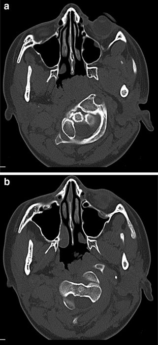
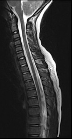
Results: Epidemiology, initial management at the scene of injury, radiological findings and pitfalls of cervical spine trauma are outlined. Strategies for conservative and surgical management are detailed depending on the pattern of injury. The management of spinal cord injuries without radiological abnormality (SCIWORA) and cranio-cervical arterial injuries is also reviewed.

Conclusions: Due to a paucity of evidence in these rare conditions, expert opinion is necessary to guide best practice management and to ensure the best chance of a good outcome for the injured child.

Keywords: Cervical spine; Children; Fracture; Pediatric; SCIWORA; Trauma.


Conflict of interest statement

All authors declare that they have no conflict of interest.

Figures

Fig. 1
Fig. 1
X-ray demonstrating C2 on C3 pseudosubluxation
Fig. 2
Fig. 2
CT images demonstrating synchondroses of: a C1 on axial view; b C2 on axial view; and c the subaxial spine on coronal view
Fig. 3
Fig. 3
CT showing atlanto-occipital dislocation: a coronal; and b sagittal. Case courtesy of Dr Yair Glick, Radiopaedia.org, rID: 52910
Fig. 4
Fig. 4
CT example of orthotopic Os Odontoideum. a Sagittal; and b coronal view. Case courtesy of Dr Roberto Schubert, Radiopaedia.org, rID: 14258
Fig. 5
Fig. 5
a, b Axial CT images demonstrating rotatory subluxation of C1 on C2. The left lateral mass is located anterior and lateral to its normal position and the right lateral mass is located posteriorly and medially
Fig. 6
Fig. 6
Sagittal T2-weighted MR image showing SCIWORA. Note the hyperintensity of cord oedema following trauma to the cervical spine

References

    1. Patel JC, Tepas JJ, III, Mollitt DL, Pieper P. Pediatric cervical spine injuries: defining the disease. J Pediatr Surg. 2001;36(2):373–376. doi: 10.1053/jpsu.2001.20720. - DOI - PubMed
    1. Baumann F, Ernstberger T, Neumann C, Nerlich M, Schroeder GD, Vaccaro AR, et al. Pediatric cervical spine injuries: a rare but challenging entity. J Spinal Disord Tech. 2015;28(7):E377-84. doi: 10.1097/BSD.0000000000000307. - DOI - PubMed
    1. Vogel LC. Unique management needs of pediatric spinal cord injury patients: etiology and pathophysiology. J Spinal Cord Med. 1997;20(1):10–13. doi: 10.1080/10790268.1997.11719445. - DOI - PubMed
    1. Huisman TA, Phelps T, Bosemani T, Tekes A, Poretti A. Parturitional injury of the head and neck. J Neuroimaging. 2015;25(2):151–166. doi: 10.1111/jon.12144. - DOI - PubMed
    1. Mortazavi M, Gore PA, Chang S, Tubbs RS, Theodore N. Pediatric cervical spine injuries: a comprehensive review. Childs Nerv Syst. 2011;27(5):705–717. doi: 10.1007/s00381-010-1342-4. - DOI - PubMed

MeSH terms

LinkOut - more resources