

T cell receptor sequencing of activated CD8 T cells in the blood identifies tumor-infiltrating clones that expand after PD-1 therapy and radiation in a melanoma patient
- PMID: 30167863
- PMCID: PMC6196100
- DOI: 10.1007/s00262-018-2228-7
T cell receptor sequencing of activated CD8 T cells in the blood identifies tumor-infiltrating clones that expand after PD-1 therapy and radiation in a melanoma patient
Abstract
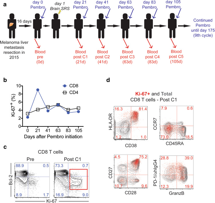
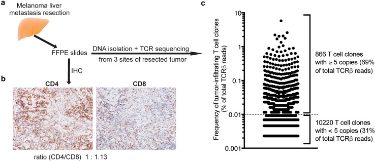
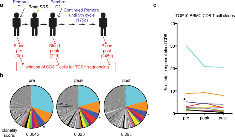
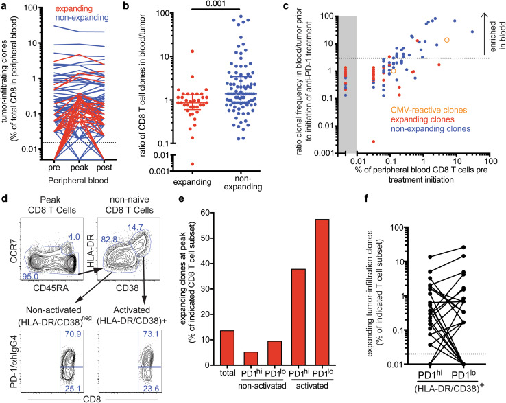
PD-1-targeted therapy has dramatically changed advanced cancer treatment. However, many questions remain, including specificity of T cells activated by PD-1 therapy and how peripheral blood analysis correlates to effects at tumor sites. In this study, we utilized TCR sequencing to dissect the composition of peripheral blood CD8 T cells activated upon therapy, comparing it with tumor-infiltrating lymphocytes. We report on a nonagenarian melanoma patient who showed a prominent increase in peripheral blood Ki-67 + CD8 T cells following brain stereotactic radiation and anti-PD-1 immunotherapy. Proliferating CD8 T cells exhibited an effector-like phenotype with expression of CD38, HLA-DR and Granzyme B, as well as expression of the positive costimulatory molecules CD28 and CD27. TCR sequencing of peripheral blood CD8 T cells revealed a highly oligoclonal repertoire at baseline with one clonotype accounting for 30%. However, the majority of dominant clones-including a previously identified cytomegalovirus-reactive clone-did not expand following treatment. In contrast, expanding clones were present at low frequencies in the peripheral blood but were enriched in a previously resected liver metastasis. The patient has so far remained recurrence-free for 36 months, and several CD8 T cell clones that expanded after treatment were maintained at elevated levels for at least 8 months. Our data show that even in a nonagenarian individual with oligoclonal expansion of CD8 T cells, we can identify activation of tumor-infiltrating CD8 T cell clones in peripheral blood following anti-PD-1-based immunotherapies.
Keywords: CD8 T cells; Immunotherapy; Melanoma; PD-1; T cell repertoire.
Conflict of interest statement
Rafi Ahmed is an inventor on patents held by Emory University that cover the topic of PD-1-directed immunotherapy. All other authors declare no potential conflicts of interest.
Figures
Similar articles
-
Costimulation through the CD137/4-1BB pathway protects human melanoma tumor-infiltrating lymphocytes from activation-induced cell death and enhances antitumor effector function.J Immunother. 2011 Apr;34(3):236-50. doi: 10.1097/CJI.0b013e318209e7ec. J Immunother. 2011. PMID: 21389874 Free PMC article.
-
PD-1+ Polyfunctional T Cells Dominate the Periphery after Tumor-Infiltrating Lymphocyte Therapy for Cancer.Clin Cancer Res. 2017 Oct 1;23(19):5779-5788. doi: 10.1158/1078-0432.CCR-16-1692. Epub 2017 Jul 5. Clin Cancer Res. 2017. PMID: 28679768 Free PMC article. Clinical Trial.
-
Metastatic Melanoma Patient Had a Complete Response with Clonal Expansion after Whole Brain Radiation and PD-1 Blockade.Cancer Immunol Res. 2017 Feb;5(2):100-105. doi: 10.1158/2326-6066.CIR-16-0223. Epub 2017 Jan 6. Cancer Immunol Res. 2017. PMID: 28062513 Free PMC article.
-
Recent Advances in Targeting CD8 T-Cell Immunity for More Effective Cancer Immunotherapy.Front Immunol. 2018 Jan 22;9:14. doi: 10.3389/fimmu.2018.00014. eCollection 2018. Front Immunol. 2018. PMID: 29403496 Free PMC article. Review.
-
A Systematic Review of the Tumor-Infiltrating CD8+ T-Cells/PD-L1 Axis in High-Grade Glial Tumors: Toward Personalized Immuno-Oncology.Front Immunol. 2021 Sep 17;12:734956. doi: 10.3389/fimmu.2021.734956. eCollection 2021. Front Immunol. 2021. PMID: 34603316 Free PMC article.
Cited by
-
Single-Cell Approaches to Profile the Response to Immune Checkpoint Inhibitors.Front Immunol. 2020 Mar 20;11:490. doi: 10.3389/fimmu.2020.00490. eCollection 2020. Front Immunol. 2020. PMID: 32265933 Free PMC article. Review.
-
Immune-awakening revealed by peripheral T cell dynamics after one cycle of immunotherapy.Nat Cancer. 2020 Feb;1(2):210-221. doi: 10.1038/s43018-019-0022-x. Epub 2020 Feb 10. Nat Cancer. 2020. PMID: 32110781 Free PMC article.
-
Modulation of CD8+ T Cell Responses by Radiotherapy-Current Evidence and Rationale for Combination with Immune Checkpoint Inhibitors.Int J Mol Sci. 2023 Nov 24;24(23):16691. doi: 10.3390/ijms242316691. Int J Mol Sci. 2023. PMID: 38069014 Free PMC article. Review.
-
Recirculation and Residency of T Cells and Tregs: Lessons Learnt in Anacapri.Front Immunol. 2020 May 5;11:682. doi: 10.3389/fimmu.2020.00682. eCollection 2020. Front Immunol. 2020. PMID: 32431695 Free PMC article. Review.
-
T-cell receptor determinants of response to chemoradiation in locally-advanced HPV16-driven malignancies.Front Oncol. 2024 Jan 3;13:1296948. doi: 10.3389/fonc.2023.1296948. eCollection 2023. Front Oncol. 2024. PMID: 38234396 Free PMC article.
References
-
- Weber J, Mandala M, Del Vecchio M, Gogas HJ, Arance AM, Cowey CL, Dalle S, Schenker M, Chiarion-Sileni V, Marquez-Rodas I, Grob JJ, Butler MO, Middleton MR, Maio M, Atkinson V, Queirolo P, Gonzalez R, Kudchadkar RR, Smylie M, Meyer N, Mortier L, Atkins MB, Long GV, Bhatia S, Lebbe C, Rutkowski P, Yokota K, Yamazaki N, Kim TM, de Pril V, Sabater J, Qureshi A, Larkin J, Ascierto PA, CheckMate C. Adjuvant Nivolumab versus Ipilimumab in Resected Stage III or IV Melanoma. N Engl J Med. 2017;377(19):1824–1835. doi: 10.1056/NEJMoa1709030. - DOI - PubMed
-
- Goldberg SB, Gettinger SN, Mahajan A, Chiang AC, Herbst RS, Sznol M, Tsiouris AJ, Cohen J, Vortmeyer A, Jilaveanu L, Yu J, Hegde U, Speaker S, Madura M, Ralabate A, Rivera A, Rowen E, Gerrish H, Yao X, Chiang V, Kluger HM. Pembrolizumab for patients with melanoma or non-small-cell lung cancer and untreated brain metastases: early analysis of a non-randomised, open-label, phase 2 trial. Lancet Oncol. 2016;17(7):976–983. doi: 10.1016/S1470-2045(16)30053-5. - DOI - PMC - PubMed
-
- Parakh S, Park JJ, Mendis S, Rai R, Xu W, Lo S, Drummond M, Rowe C, Wong A, McArthur G, Haydon A, Andrews MC, Cebon J, Guminski A, Kefford RF, Long GV, Menzies AM, Klein O, Carlino MS. Efficacy of anti-PD-1 therapy in patients with melanoma brain metastases. Br J Cancer. 2017;116(12):1558–1563. doi: 10.1038/bjc.2017.142. - DOI - PMC - PubMed
-
- Schadendorf Dirk, Ascierto Paolo Antonio, Haanen John B. A. G., Espinosa Enrique, Demidov Lev V., Garbe Claus, Lorigan Paul, Gogas Helen, Hoeller Christoph, Guren Tormod Kyrre, Rorive Andree, Rutkowski Piotr, Muñoz-Couselo Eva, Dummer Reinhard, Carneiro Ana, Hospers Geke, Grigoryeva Elena Borisovna, Bhore Rafia, Nathan Paul. Efficacy and safety of nivolumab (NIVO) in patients with advanced melanoma (MEL) and poor prognostic factors who progressed on or after ipilimumab (IPI): Results from a phase II study (CheckMate 172) Journal of Clinical Oncology. 2017;35(15_suppl):9524–9524. doi: 10.1200/JCO.2017.35.15_suppl.9524. - DOI
Publication types
MeSH terms
Substances
Grants and funding
LinkOut - more resources
Full Text Sources
Other Literature Sources
Medical
Research Materials
